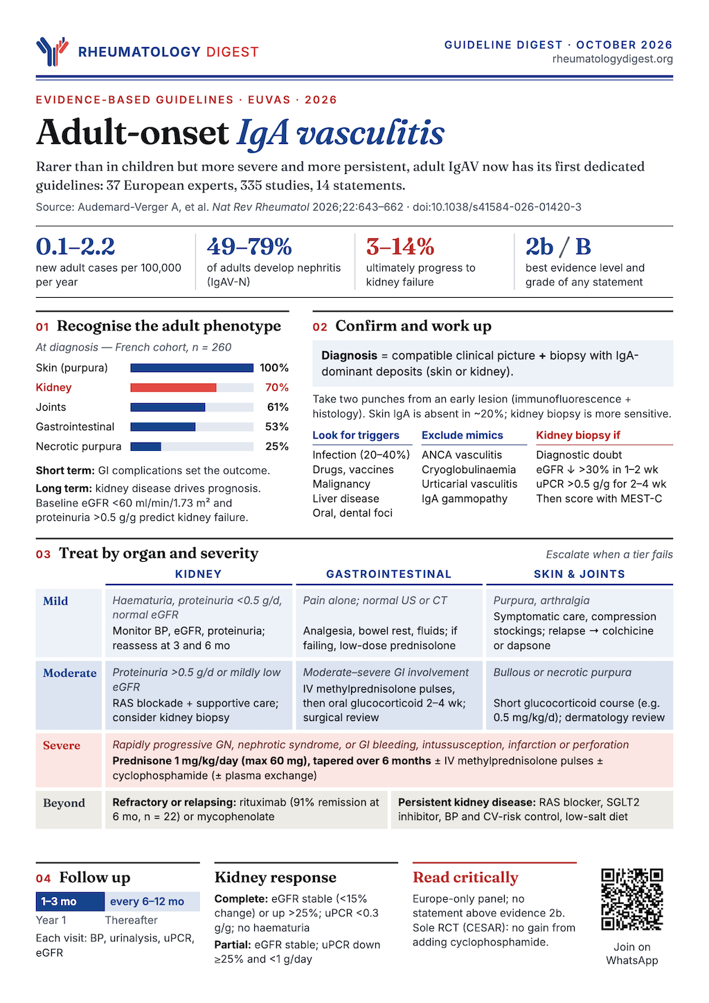
TL;DR: The first guidelines for adult IgA vasculitis give a practical framework — confirm with histology, screen for triggers and malignancy, treat by organ and severity, and watch the kidney for years — but rest mostly on observational data and expert opinion.
IgA vasculitis in adults has long been managed by borrowing from children, where the disease is common and usually self-limiting, and from IgA nephropathy, where the kidney lesion looks the same. This guideline from the European Vasculitis Society is the first to be written for adults. It is a consensus document built on a systematic review, and its value lies as much in what it admits is unknown as in what it recommends.
How the guideline was built
The group followed the RIGHT reporting framework. A core committee of nephrologists, dermatologists, rheumatologists and a methodologist worked with a voting panel, giving 37 experts in total, including a pathologist, paediatric, internal medicine and vasculitis nurse specialists, and a patient representative. Of 21 initial clinical questions, 14 reached consensus in a two-round Delphi process and were converted into PICO questions.
The literature search ran from 1970 to November 2025 across PubMed, Embase and Cochrane. It identified 3,784 articles, of which 335 informed the guideline. Studies of children were considered only when adult evidence was missing. Evidence and recommendations were graded on the Oxford scheme, and a statement was accepted if more than 80% of the panel scored agreement at 8 out of 10 or higher. Mean agreement across the 14 statements ran from 8.8 to 9.7.
The disease in adults
IgAV, formerly Henoch–Schönlein purpura, is an immune complex-mediated small-vessel vasculitis with IgA deposition in vessel walls as its pathological hallmark. It typically involves the skin, gut, kidneys and joints. Incidence is about 3–27.2 per 100,000 in children and 0.1–2.2 per 100,000 in adults. Paediatric disease is often a single episode and self-limiting, whereas adult disease is more severe, more persistent and associated with a poorer prognosis.
The typical presentation is purpura of the lower limbs, often preceding other features. Joint symptoms, abdominal pain (colicky pain and gastrointestinal bleeding) and kidney disease follow less often. Involvement of other organs, including lungs, heart and testes, is rare in adults.
Kidney disease is the long-term determinant. IgAV with nephritis (IgAV-N) is histologically indistinguishable from IgA nephropathy: both have IgA-dominant glomerular deposits with mesangial and endocapillary hypercellularity and extracapillary proliferation, and both involve galactose-deficient IgA1 and its antibodies. The guideline notes differences too. IgAV-N shows more endocapillary hypercellularity and crescents, more extensive capillary-wall deposition of IgA, IgG and C3, and less chronicity than IgAN, in which glomerulosclerosis and tubular atrophy are more common. Kidney involvement is reported in 49–79% of adults, although this may be overestimated, since most cohorts were recruited by rheumatology or nephrology centres that see more systemic disease. About 29% experience worsening, and 3–14% ultimately develop kidney failure. In one French cohort followed for a median of 14.8 years, 38% developed CKD and 11% progressed to kidney failure. Rates of CKD are much higher in adults than children.
Diagnosis: histology is expected, and IgA may be missing
Statement 1. Diagnosis should rest on clinical, laboratory and histological findings together. Predominant IgA deposits with compatible histology (such as mesangioproliferative glomerulonephritis or cutaneous leukocytoclastic vasculitis) in a patient with a suggestive picture strongly supports the diagnosis, but the absence of IgA deposits does not exclude it. (Level 4, grade C.)
The practical points in the text are specific:
- Skin biopsy is accessible and shows leukocytoclastic vasculitis, which is not specific. Direct immunofluorescence is the key test, and typically shows granular IgA in or around vessel walls.
- Timing matters. IgA may degrade in older lesions, so biopsy early lesions, which look like partly blanching macules with minor petechial and papular components, often next to palpable purpura. Ideally take two punch biopsies: one for immunofluorescence (not in formalin) and one for routine histology.
- Perivascular IgM or IgG without IgA should prompt a search for other diagnoses, including cryoglobulinaemic vasculitis, rheumatoid vasculitis, Sjögren disease, lupus vasculitis, hypocomplementaemic urticarial vasculitis and AAV.
- A negative skin result does not end the question. In a French cohort of 260 adults, 20% had no detectable IgA in skin biopsies, whereas IgA was found in kidney biopsy specimens, so a kidney biopsy showing IgA can establish the diagnosis. Kidney biopsy appears more sensitive than skin biopsy for detecting IgA.
Statement 2. Classification. No criteria were developed specifically for adults. The panel supports using the EULAR/PRINTO/PReS Ankara 2008 criteria, developed in children, for adults too, after excluding AAV and cryoglobulinaemic vasculitis, but only to classify patients for clinical studies, not for diagnosis. (Level 2b, grade B.) The older 1990 ACR criteria included age under 20 and granulocytic infiltration, which limits their use in adults. A study found Ankara superior to ACR in adults, and its performance improved further when ANCA positivity or cryoglobulins were treated as exclusions: sensitivity 76–97.8% and specificity 85.0–94.5% across two cohorts.
The work-up: triggers, mimics and associated disease
Statement 3. At diagnosis, look for triggers and associated conditions: acute infections, newly introduced drugs or vaccines, age- and sex-appropriate screening for solid and haematological malignancy, chronic infections (such as viral hepatitis or oral and dental disease), and liver disease. (Level 2b, grade B.) The text adds detail:
- Infection precedes IgAV in about 20–40%, typically bacterial infection of the respiratory, urinary or gastrointestinal tract. For dental sources, adult data are lacking, but evaluation could be considered.
- Drugs. A pharmacovigilance study using WHO’s VigiBase found vaccines, antibiotics and TNF inhibitors were the drug classes most often associated. Cases after SARS-CoV-2 infection or COVID-19 vaccination have been reported, but causality cannot be established.
- Malignancy. A systematic review identified over 100 published cases of malignancy-associated IgAV, 67% involving solid tumours and 33% haematological. Lung cancer was most common among solid tumours, followed by genitourinary and gastrointestinal. Plasma cell dyscrasia accounted for 43% of the haematological cases. In a prospective study, 7.5% of adults had cancer, most diagnosed within three years either side of IgAV. In a separate retrospective study of 30 patients with malignancy-associated IgAV, those with cancer were older than patients without it and more often had severe features such as necrotic purpura and alveolar haemorrhage.
- Liver disease. A few reports link IgAV to cirrhosis, mostly alcohol-related, and the guideline recommends systematic screening for occult liver disease, particularly with excessive alcohol use.
The paper’s diagnostic work-up (their Figure 3) groups the essential investigations into microbiology (HCV, HBsAg, HIV and cultures), malignancy and associated disease screening (including screening for IgA monoclonal gammopathy, blood count and film, LDH, biochemistry and liver tests), organ assessment (skin biopsy with IgA staining, abdominal ultrasound or CT, and kidney assessment by urine dipstick, urinary protein-to-creatinine ratio, creatinine and eGFR), and immunology (ANA, ANCA, immunoglobulins, C3 and C4, cryoglobulins and rheumatoid factor).
Gastrointestinal involvement
Statement 4. Abdominal pain is very common. Massive bleeding, intussusception, intestinal infarction or perforation define severe gastrointestinal involvement, which is potentially life-threatening and needs urgent management. (Level 2b, grade C.)
Gastrointestinal involvement affects 13–60% of adults. In a large Spanish series the main manifestations were abdominal pain, nausea and vomiting, and melaena or haematochezia; in the French multicentre cohort 53% had involvement, with abdominal pain in 99%. Mesenteric ischaemia and perforation were fatal in some. Faecal occult blood testing can support the diagnosis, the descending duodenum and terminal ileum are often involved, and endoscopy shows erythema, petechiae, haemorrhagic erosions and ulcers. Biopsies typically show inflammatory infiltrates and erythrocyte extravasation and often no vasculitis, so their diagnostic yield is limited and they carry a risk of perforation. Adults more often get CT than ultrasound, and abdominal CT is abnormal in about 60% of those with suspected involvement, most commonly with bowel wall thickening or mesenteric vessel engorgement.
Kidney involvement and when to biopsy
Statement 5. Suspect kidney involvement in patients with haematuria, proteinuria or reduced eGFR. Severe presentations are nephrotic syndrome and rapidly progressive glomerulonephritis (RPGN). An eGFR below 60 ml/min/1.73 m² and proteinuria above 0.5 g/g are the main baseline factors associated with progression to kidney failure. (Level 2b, grade B.)
Haematuria (more than five red cells per high-power field), proteinuria above 300 mg/day or ACR above 30 mg/g, or reduced eGFR during the course should prompt suspicion. Microscopic haematuria is present in almost all patients with IgAV-N, with subnephrotic proteinuria common and about a third having impaired kidney function. Nephrotic syndrome occurs in up to 23% and RPGN with diffuse crescentic disease in about 10%. Abnormalities can appear weeks or months after the first signs, so adults with new IgAV should have regular urinalysis and kidney function checks. Other factors linked to poor outcome, in some but not all studies, are hypertension and age over 50.
Statement 6. Biopsy is indicated when there is diagnostic uncertainty about IgAV-N in a patient with systemic vasculitis, or when kidney function worsens rapidly (a decline above 30% in eGFR over 1–2 weeks) or proteinuria stays above 0.5 g/g for 2–4 weeks on or off treatment. Kidney involvement should be classified using the Oxford MEST-C score. (Level 2b, grade C.) The reported complication risk is low: bleeding needing transfusion in 1.6% and interventions to control bleeding in 0.3%.
On classification, no histological scheme was designed for adult IgAV-N. Several studies used the paediatric ISKDC grading, which is largely based on the proportion of glomeruli with crescents, but most adult biopsies are grade II or III and in a French cohort of 250 adults crescent proportion did not independently predict outcome. The MEST-C score was evaluated in a cohort of 99 adults and 262 children: endocapillary hypercellularity (E1) was associated with a high-risk eGFR trajectory, with an early improvement followed by a later decline 3–4 years after biopsy, and patients with E1 lesions need long-term monitoring. A 2026 meta-analysis supported the prognostic value of E1 and T lesions. Because most patients in these cohorts received immunosuppression, the predictive value of lesions is hard to disentangle from treatment.
Treatment: the goal, then the organ
Statement 7. Treatment aims for clinical remission and prevention of damage, especially kidney damage, and should reflect disease severity and comorbidities. Options run from supportive care to glucocorticoids, with or without immunosuppressive or immunomodulatory agents. Underlying diseases (such as neoplasms) should be treated and potential triggers (such as infections) eliminated. (Level 2b, grade B.) Evidence comes mostly from observational studies and a few small trials. Remission is generally defined as a Birmingham Vasculitis Activity Score of 0.
The four overarching principles are to aim for quality of life while preventing relapse, preserving organ function and improving survival; to give patients the highest standard of care, preferably at or with a vasculitis centre; to manage the disease through an interdisciplinary team with shared decision-making; and to make patient education an ongoing pillar of care.
The paper’s treatment algorithm (their Figure 4) tiers kidney involvement into mild, moderate and severe. Mild (haematuria, proteinuria below 0.5 g/day, normal eGFR) means monitoring blood pressure, eGFR and proteinuria and reassessing at 3 and 6 months. Moderate (proteinuria above 0.5 g/day, mildly reduced eGFR) means RAS blockade and supportive therapy, with kidney biopsy considered. Severe (RPGN or nephrotic syndrome) means prednisone 1 mg/kg/day (maximum 60 mg) tapered over 6 months, with or without intravenous methylprednisolone pulses, cyclophosphamide and plasma exchange, followed by B-cell depletion (rituximab) or MMF if refractory or relapsing.
Skin
Statement 8. No skin-specific treatment is needed when glucocorticoids or immunosuppressants are being given for systemic disease. Colchicine or dapsone may be considered for relapses limited to the skin, and compression stockings may relieve lower-leg purpura. For skin-limited disease with haemorrhagic bullous or necrotic purpura, short glucocorticoid courses are recommended. (Level 4, grade C.)
Details: acute flares usually last days to 1–2 weeks. Compression may reduce new lesions. Systemic glucocorticoids (for example 0.5 mg/kg/day) are suggested when incipient necrosis is suspected, heralded by haemorrhagic blisters, because healing is prolonged once ulcers form; use 3–5 day courses and reassess. For flares beyond 7 days or early relapses, add colchicine (1 mg/day increasing to 1.5 mg/day after a week if tolerated) or dapsone (1–2 mg/kg/day, after excluding G6PD deficiency). Azathioprine, methotrexate and hydroxychloroquine are not advised for skin-limited disease, and colchicine or dapsone is usually given for six months before attempting withdrawal.
Gastrointestinal
Statement 9. Mild gastrointestinal disease (isolated abdominal pain with normal ultrasound or CT) needs symptomatic treatment, a light diet and close monitoring. Moderate to severe cases should be considered for glucocorticoid pulses and/or oral glucocorticoids, given as short courses when no other severe systemic manifestation exists. Plasma exchange or cyclophosphamide can be considered as salvage for refractory disease. (Level 2b, grade C.)
In practice: in a French series of 135 adults with GI involvement, 15% received no glucocorticoids and still had a favourable outcome. Severe cases usually need admission, and a fasting period with enteral or parenteral nutrition may be considered. For moderate-to-severe disease, methylprednisolone pulses (typically 3 days at 7.5–15 mg/kg/day) are often followed by oral prednisone 1 mg/kg/day for 2–4 weeks. Cyclophosphamide is a salvage option on a case-by-case basis; the CESAR randomised trial found no benefit from adding it to glucocorticoids, but only 15 patients had severe GI disease, which precludes firm conclusions.
Kidney
Statement 10. Prescribe glucocorticoids in IgAV-N when there are poor prognostic factors: impaired kidney function, nephrotic-range proteinuria, proteinuria above 0.5 g/g persisting despite RAS blockade, or RPGN. An immunosuppressant and/or plasma exchange can be discussed in RPGN. For patients reaching kidney failure, peritoneal dialysis and haemodialysis are both feasible, and transplantation usually has favourable outcomes. (Level 2b, grade B; this statement had the lowest agreement, 8.8.)
The text is candid about the evidence:
- In patients without kidney involvement at presentation, neither glucocorticoids nor other therapies appear to prevent IgAV-N later.
- Regimens are borrowed from IgAN and the 2025 KDIGO guideline. The Pozzi–Locatelli regimen combines prednisone with methylprednisolone pulses but is rarely used because of cumulative glucocorticoid exposure. The revised TESTING regimen is now preferred: oral methylprednisolone 0.4 mg/kg/day (maximum 32 mg), tapered by 4 mg/day each month over 6–9 months. The best duration is unknown, and a minimum of 3–6 months is considered reasonable.
- Steroids alone are often not enough. In the French cohort of 260 adults, complete remission was achieved in only about 30% of those treated with glucocorticoids alone and relapse occurred in 22%, though the stringent BVAS-0 definition and small numbers temper that.
- Add-on immunosuppression is a case-by-case discussion in RPGN or relapsing disease and as a steroid-sparing step. Cyclophosphamide has case series support but the only RCT (CESAR) did not show benefit. In a UK retrospective study, MMF was the second most prescribed drug after glucocorticoids; among patients who started it within 6 months of diagnosis, 6 of 13 (46%) had responded by month 6 and 6 of 11 (55%) by month 12, though no specific kidney effect could be shown. Evidence for azathioprine or calcineurin inhibitors in adults is scarce.
- Plasma exchange may be considered in IgAV-associated RPGN: in a French series of 11 severely affected patients it was followed by improved kidney function and proteinuria, but a systematic review found only four adults among 38 patients, all of whom also received steroids and other immunosuppression. The panel suggests it by analogy with AAV.
- Kidney replacement and transplantation. Choice of dialysis should follow patient preference. Peritoneal dialysis has a theoretical drawback, since GI flares could be confused with peritonitis, but there is no evidence to prefer haemodialysis in stable disease. Transplantation is recommended for kidney failure without extra-renal activity. Outcomes in comparison studies are similar to matched controls, but recurrence is a real risk: in one Italian study it was 71% in grafts of patients whose native kidneys had necrotising or crescentic disease versus 12% in those with mesangioproliferative disease, and a US registry study found recurrence in 29.7% of adults and 13.0% of children with increased graft loss.
Statement 11. Supportive care. All patients with clinically relevant persistent kidney involvement should receive lifestyle advice, cardiovascular protection, antihypertensive and antiproteinuric therapy (RAS blockers), and SGLT2 inhibitors, following KDIGO guidance for glomerular disease. (Level 2b, grade B.) The guideline notes that patients with mild skin-limited disease and only transient or mild haematuria or proteinuria are unlikely to need the full package. SGLT2 inhibitors have not been studied extensively in IgAV-N, but the recommendation extrapolates from KDIGO advice for IgAN at risk of progressive loss of kidney function.
Biologics and what is coming
Statement 12. Biologics, particularly rituximab, can be considered in relapsing or refractory adult IgAV. (Level 4, grade C.) Evidence is limited to case reports and series, and the rationale is to target cells producing abnormal IgA1 and anti-IgA1 antibodies.
- Italian observational study, 22 adults (relapsing/refractory disease or contraindications to standard drugs): remission in 91% at 6 months, with one patient progressing to kidney failure. Kidney function remained stable and proteinuria fell over a median of 24 months. Fifteen (68%) had kidney involvement and 9 had diffuse endocapillary or extracapillary glomerulonephritis.
- Twelve adults with severe glomerulonephritis (all with extracapillary proliferation): complete response in 11 (92%), mostly with rituximab as monotherapy.
- Systematic review of 35 treated cases (paediatric and adult): almost 90% had kidney involvement, and improvement occurred in 94%, sustained remission in 74%. Of 13 relapses, 11 were retreated successfully. Rituximab-related adverse events occurred in 8.6%, mostly minor, with no deaths.
- Target trial emulation, 368 patients comparing rituximab with cyclophosphamide: 35% lower risk of kidney failure or CKD stage IV–V with rituximab, with the possibility of indication bias.
- An RCT of rituximab versus placebo in severe adult IgAV (NCT05329090) is ongoing, including patients with severe GI disease.
Other agents (ofatumumab, bortezomib and telitacicept, the last in a pilot of 13 adults) are anecdotal. TNF inhibitors and IL-1 inhibitors are routinely given for coexisting spondyloarthritis, IBD or familial Mediterranean fever, but IgAV has been reported after TNF inhibitors and their effect on IgAV is unclear. New IgAN therapies, including targeted-release budesonide, sibeprenlimab (a B-cell-activating factor-APRIL axis inhibitor) and iptacopan, may be relevant to IgAV-N, and the authors say the trials largely excluded IgAV-N, so they warrant investigation.
Measuring disease, remission and relapse
Statement 13. There is no IgAV-specific activity measure. The Birmingham Vasculitis Activity Score can be used as a generic tool, though a disease-specific one would be preferable. Remission is the absence of any active disease manifestation; response is clinically meaningful improvement of known manifestations without new ones; relapse is recurrence or new onset of disease-related manifestations after remission or response. (Level 5, grade C.)
The reasons BVAS fits poorly are specific: several domains it assesses in detail (eye, ear–nose–throat) are rarely involved in IgAV, and in the kidney domain it weights haematuria more heavily than proteinuria, whereas proteinuria is the stronger predictor of kidney outcome in IgAN and IgAV-N and haematuria is a common feature not clearly tied to progression. The panel proposes kidney definitions, which are arbitrary and need validation:
- Complete remission: stable kidney function (eGFR variation below 15%) if baseline was normal, or an eGFR improvement above 25% if baseline was impaired by IgAV-N, plus a urinary protein-to-creatinine ratio below 0.3 g/g and no haematuria.
- Partial remission: stable kidney function (variation below 15%) and a reduction of at least 25% in UPCR from peak, with an absolute value below 1 g/day.
Follow-up
Statement 14. Adult IgAV can follow a chronic–relapsing course. Follow-up duration should be tailored to the baseline presentation, its severity and relapses. The recommendation is to see patients every 1–3 months for the first year and every 6–12 months thereafter, with tests that detect kidney involvement in good time. (Level 5, grade C.) The frequency is acknowledged to be largely expert consensus, to be tailored to the course, and no consensus exists on optimal kidney monitoring.
Relapse rates vary widely between studies. Some patients relapse at least once, most often in skin, joints or kidneys, and gastrointestinal relapses are uncommon. At relapse, all organs potentially affected should be evaluated, and regular laboratory checks should continue even when disease is quiet. Persistent urinary abnormalities are common even after treatment, and proteinuria during follow-up is a key risk factor for progression. Hypertension, diabetes and pre-existing CKD may worsen outcome. The survival data in the paper’s tables show the stakes: in one French cohort, 25.6% of 250 patients had died by follow-up, while a UK analysis of 2,838 patients found all-cause mortality 8.4% and a hazard ratio of 1.3 for death and 1.5 for CKD stages 3–5.
What the guideline cannot settle
The authors name the limits themselves:
- The evidence is thin. The highest level reached was 2b and the highest grade B, reflecting the scarcity of randomised trials and meta-analyses in adults. Many statements are level 4–5 and grade C.
- Geography. Representation was restricted to Europe, which may limit generalisability.
- Some recommendations are admittedly expert opinion, such as the follow-up schedule and the kidney remission definitions.
- IgAV-N regimens are extrapolated from IgAN, with unclear optimal duration and an unproven benefit of immunosuppression beyond steroids.
Their research agenda calls for diagnostic criteria for adults, randomised trials in IgAV-N (including repurposed IgAN therapies, complement inhibitors and novel biologics), disease-activity biomarkers, an IgAV-specific activity score, validation of remission, response, relapse and refractory-disease definitions, a clinicopathological scoring system to guide treatment, and precision-medicine approaches. Regular updates are planned.
What it adds up to
Three practical messages stand out. First, the diagnosis is a team exercise: get tissue, take two punch biopsies of early lesions, and remember that missing IgA does not exclude IgAV while a kidney biopsy may succeed where skin failed. Second, adults need a hunt for causes that children do not: drugs, infections, liver disease and especially malignancy, with plasma cell dyscrasia a recurring one. Third, the kidney sets the long-term course, so the follow-up that matters is repeated urine and eGFR checks for years, with proteinuria above 0.5 g/g and eGFR below 60 as the early warning signs.
What the guideline does not offer is a proven treatment algorithm. Steroids dominate, add-on immunosuppression rests on small series and one negative RCT, and the case for rituximab rests on promising but uncontrolled data, with a trial under way.
Disclosures noted in the source: many panel members report consulting, speaker and research relationships with pharmaceutical companies, listed in full in the paper.