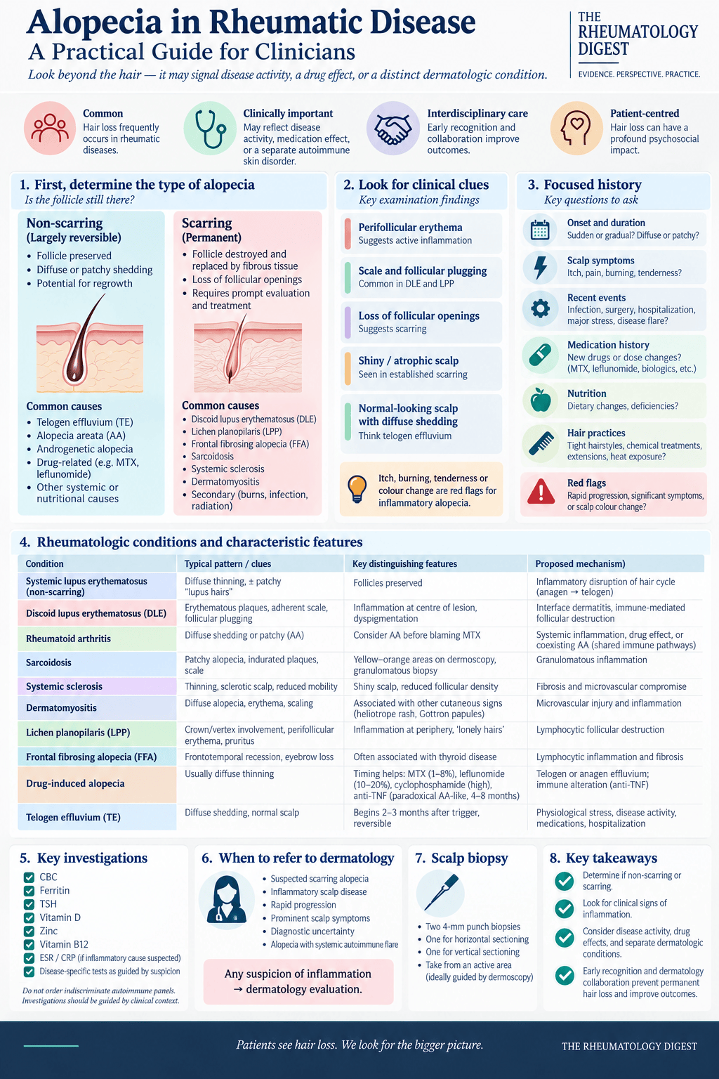
TL;DR: In rheumatic disease the first question about hair loss is not which disease caused it, but whether it is scarring — that single distinction decides whether the follicle can still be saved.
Hair loss is one of the complaints most likely to be waved through in a rheumatology clinic. It rarely features in a disease activity score, it is not in the core set, and the reflexive answer — “it’s the methotrexate, it’ll settle” — is right often enough to be dangerous. This review, from a dermatology group writing for rheumatologists, argues that the reflex costs follicles. Alopecia in rheumatic disease can be a marker of active systemic inflammation, an adverse drug effect, a separate autoimmune comorbidity, or an irreversible scarring process that is quietly destroying stem cells while the patient waits for a dermatology appointment. The four are managed very differently, and only one of them forgives delay.
Start with the fork that decides everything
The hair follicle cycles between anagen (growth), catagen (apoptosis-mediated regression) and telogen (quiescence), under the control of immune signalling, hormones, vascular supply and environmental stress. Anything that disrupts that regulation, or damages the follicle directly, produces hair loss — but the clinically decisive question is which.
Non-scarring alopecia leaves the follicle intact. The hair cycle is disrupted or the follicle is transiently inflamed, and regrowth remains possible once the underlying cause is addressed. Scarring (cicatricial) alopecia destroys the follicular epithelium and its stem cells, replacing the follicle with fibrous tissue. That loss is permanent. Everything about prognosis, urgency and management flows from this single distinction, which is why the review puts it before any disease-specific discussion.
The scalp examination is where the fork is usually resolved. Erythema, scale, follicular plugging and — most importantly — loss of follicular openings point to a permanent process. Perifollicular erythema or induration is the red flag that ongoing inflammation is actively threatening follicular integrity. Symptoms help too: pruritus, pain, burning or tenderness suggest an inflammatory or scarring process, whereas telogen effluvium and alopecia areata are typically asymptomatic.
This is also not a cosmetic aside. Hair is tied to identity, self-confidence and cultural norms, and patients with alopecia report reduced self-esteem, social withdrawal, embarrassment, perceived stigmatisation and measurable decline in quality of life. Anxiety and depression are more prevalent in patients with alopecia than in controls — and in rheumatic disease that burden sits on top of chronicity, physical limitation and the adverse effects of immunomodulatory therapy.
Telogen effluvium: the default explanation, and how to confirm it
Telogen effluvium (TE) is diffuse, non-scarring shedding caused by a disruption in hair cycling, and it is the pattern rheumatologists see most. Normally around 90% of scalp hairs are in anagen and 10% in telogen. A stressor drives a large cohort of anagen hairs into premature telogen; when the new anagen hair pushes the dormant club hair out, the patient sheds. The shedding typically starts two to three months after the trigger and is self-limiting once the cause is removed — but the volume can be dramatic enough to frighten both patient and clinician.
Patients with rheumatic disease are predisposed on several fronts at once: chronic systemic inflammation, elevated circulating cytokines, metabolic stress, acute disease flares, hospitalisations, surgery, and the antimetabolites and immunomodulators used to treat them.
The confirmatory findings are reassuringly simple. Shedding is diffuse rather than patchy — hair fall, not discrete patches. The hair pull test is positive. Crucially, the scalp itself looks normal: no erythema, no perifollicular scale, and follicular openings preserved. Trichoscopy shows reduced density and increased empty follicular units. Histology, if obtained, shows an increased proportion of telogen hairs (>25%) without significant inflammation.
Getting this right matters in both directions. Recognising TE tells you to look for the physiological stressor or the disease flare, rather than chasing primary follicular pathology — and recognising that it is not TE is what stops a scarring alopecia being reassured away.
Lupus has two alopecias, and they behave in opposite ways
Reported prevalence of alopecia in SLE ranges from 17.3% to 85.2% — a spread wide enough to be its own finding, and one the authors flag as unresolved.
The diffuse, non-scarring form is the common one: widespread thinning driven by systemic inflammation and abnormal cycling. Immune complex deposition and complement activation in the skin and follicles create an inflammatory environment that promotes premature anagen-to-telogen transition, so shedding tracks periods of disease activity. Chronic inflammation also damages follicular keratinocytes and impairs hair shaft integrity, producing the short, fragile, dry, coarse hairs along the frontal hairline known as lupus hairs — a dishevelled appearance reflecting cumulative injury to the follicular epithelium rather than follicle destruction.
The scarring form belongs to discoid lupus erythematosus (DLE), which can occur with or without underlying SLE. Roughly 60% of DLE patients have scalp involvement, and about a third of those develop scarring alopecia. DLE produces interface dermatitis — lymphocytes attacking the dermal-epidermal junction — with lymphocytes also migrating around the follicles. The perifollicular inflammation damages the follicular structure over time: scale and follicular plugging first, then follicle collapse, disappearance of the sebaceous gland, and fibrosis. Once the inflammation reaches and destroys the stem cell reservoir, the hair cannot grow back.
Clinically the lesions begin as erythematous or violaceous plaques with scale and follicular plugging, and end as smooth scarred patches with conspicuous loss of follicular openings. Dermoscopy shows follicular keratotic plugs, telangiectasia and decreased follicular density. Histopathology shows interface dermatitis with lymphocytic infiltrate at the dermal-epidermal junction and follicular epithelium, basement membrane thickening, mucin deposition and perifollicular inflammation.
The practical instruction is the same one that runs through the whole review: identify and treat the cutaneous lupus early, because the window in which the follicle can still be saved closes. And in the other direction — unexplained alopecia with systemic symptoms or photosensitivity warrants evaluation for underlying autoimmune disease.
Rheumatoid arthritis: shedding, drugs, and a genuine alopecia areata link
In RA, hair loss most often means diffuse shedding from systemic inflammation or from medication. But the review makes a case for a third possibility that is easy to miss: alopecia areata (AA) as a comorbidity of RA, supported by shared genetic susceptibility rather than coincidence.
AA is non-scarring, and its mechanism is worth understanding because it explains both the clinical appearance and the emerging treatments. The anagen follicle is normally an immune-privileged site, protected by reduced antigen presentation, immunosuppressive signals (TGF-β, IL-10, α-MSH) and limited T-cell and NK cell infiltration. In AA that privilege collapses: MHC class I expression increases, cytotoxic T-cells are recruited, and autoreactive CD8+ T lymphocytes attack the follicular keratinocytes and hair matrix cells responsible for producing the shaft. The MHC upregulation is induced by IFN-γ acting through the JAK-STAT pathway — which is precisely why JAK inhibition has become relevant here. The follicle exits the growth phase prematurely and produces patchy loss, but the follicle itself is not destroyed, so the hair can regrow.
The overlap with RA runs through shared inflammatory networks — autoreactive T-cells and TNF-α, IL-6 and IFN-γ mediate both — and through shared susceptibility loci in the HLA region on chromosome 6. The TRAF1-C5 region, implicated in RA susceptibility and severity, has also been implicated in AA.
Clinically AA ranges from diffuse to well-demarcated patchy loss. Patches are usually asymptomatic, though tingling or itching may precede onset. Gross examination shows round or oval patches without scale or skin thickening. Dermoscopy reveals exclamation point hairs, identified by their narrowed root. Disease can progress to alopecia totalis (whole scalp) or alopecia universalis (all body hair). Biopsy shows a dense lymphocytic infiltrate around the base of the follicle — the “swarm of bees” — with pigmentary incontinence of the dermal papillae and deterioration of the bulb epithelium.
Sarcoidosis: scalp involvement may be the presenting sign
Sarcoidosis-associated hair loss occurs where the scalp is cutaneously involved, and it is characteristically patchy and scarring. Skin involvement in sarcoidosis has a reported prevalence of 24–40%, and although scalp involvement specifically is uncommon, the authors emphasise that it can be the first clinical indication of systemic disease.
The mechanism is granulomatous. Noncaseating granulomas of epithelioid histiocytes, multinucleated giant cells and lymphocytes form in the dermis, driven by dysregulated Th1 signalling with increased IFN-γ, IL-2 and TNF-α. When those granulomas develop around hair follicles and the pilosebaceous unit, the resulting perifollicular inflammation and structural destruction disrupt follicular architecture and impair the follicle’s ability to sustain anagen. In advanced disease, granulomatous damage to the follicular epithelium and stem cells produces frank cicatricial alopecia.
Clinically the scalp shows erythematous, indurated plaques with scale and localised hair loss. Trichoscopy demonstrates decreased follicular density, perifollicular scaling, and areas that appear yellow or orange — reflecting the underlying granulomatous inflammation. Histopathology is essential for confirmation, showing noncaseating epithelioid granulomas with multinucleate giant cells in the dermis.
Because scalp involvement may signal systemic inflammatory activity, it should prompt evaluation for disease elsewhere — and early treatment may prevent permanent scarring.
Scleroderma: fibrosis and perfusion, not immune destruction
Hair loss in systemic sclerosis occurs in areas of cutaneous fibrosis, and is usually a reduction in hair density rather than a discrete alopecic process. It is not among the more prominent manifestations of the disease, but it reflects the same structural changes happening in the skin.
The pathology is vascular and fibrotic. Early disease brings vascular dysfunction and chronic inflammation with fibroblast activation and infiltration of T-lymphocytes and macrophages. TGF-β and platelet-derived growth factor drive collagen deposition and dermal thickening. Within the scalp, progressive dermal fibrosis disrupts the architecture of the pilosebaceous unit, producing stiffness, reduced vascular supply to nearby tissue, and follicles that can no longer sustain normal cycling. Chronic microangiopathy compounds this by deranging follicular metabolism — blood flow to the hair bulb and matrix keratinocytes falls, and follicular activity falls with it.
The distinction the authors draw is a useful one: scleroderma-associated hair loss reflects mechanical and vascular change within fibrotic skin, not direct immune-mediated destruction of follicles. Clinically it appears as thinning or patchy alopecia within regions of indurated or sclerotic skin, and its extent offers some insight into the extent and activity of the underlying disease.
Dermatomyositis: scalp involvement that gets mistaken for seborrhoeic dermatitis
Dermatomyositis can involve the scalp with erythema, scaling, notably intense pruritus and non-scarring alopecia. The mechanism is the same microvascular injury that characterises the disease elsewhere: complement proteins deposit in the microvessels of the skin, causing damage, leakage and perivascular inflammation. The resulting microangiopathy reduces blood flow to the hair follicles, so the metabolic demands of the matrix keratinocytes cannot be met — impairing hair growth and increasing shedding. Inflammatory infiltration of the scalp adds to this, and biopsies often show interface dermatitis and perivascular lymphocytic infiltration. Chronic perifollicular inflammation induces premature transition into telogen and diffuse thinning.
The clinical trap is that scalp dermatomyositis looks like other inflammatory scalp disorders — seborrhoeic dermatitis or psoriasis. The review’s advice is to suspect it when there is persistent erythema, scaling and telangiectasia of the scalp, particularly if heliotrope rash, Gottron papules or other characteristic cutaneous findings are concurrently present. Recognising scalp involvement can bring the diagnosis forward.
Drug-induced alopecia: the conversation worth having before the first dose
Patients who lose hair on a DMARD may become reluctant to continue it, and adherence suffers. The review’s framing is pragmatic: knowing which drugs do this, by what mechanism, and over what timescale allows the conversation to happen before it becomes a reason to stop treatment.
Drug-induced alopecia is non-scarring, reversible and diffuse, and occurs by two mechanisms. Telogen effluvium typically appears two to four months after starting a drug, as follicles are sent prematurely into a resting phase. Anagen effluvium is halted hair growth from an inflammatory attack on the follicle during the growth phase itself — faster and more dramatic.
Methotrexate. As an antifolate, MTX impairs DNA synthesis in rapidly dividing cells, including the hair matrix. The reported incidence varies considerably: roughly 1–5% in a study of nearly 4,000 patients, with hair loss more common in women than men; up to 8% within a year in a 2021 study of 1,049 RA patients; but 29.4% in a 2019 study of 68 patients, 60% of them women. In that last study, patients reporting alopecia also reported a higher rate of general symptoms, pointing towards drug metabolism as the variable. That reading is supported by a case report of a patient on low-dose MTX who lost hair after just the first two doses and was found to have a genetic polymorphism in the reduced folate carrier protein responsible for intracellular MTX transport — a mutation that may raise plasma levels and, with them, risk. Higher doses, combination DMARD therapy and concomitant folate deficiency all increase risk. The preventive measure is the obvious one: consider folic acid supplementation when starting MTX.
Leflunomide. By blocking dihydroorotate dehydrogenase, leflunomide inhibits pyrimidine synthesis and interferes with rapidly dividing hair matrix cells, producing gradual thinning over weeks to months through a telogen effluvium mechanism. Drug-induced hair loss or diffuse thinning occurs in around 10–20% of patients, and the highest risk is in those on concomitant DMARDs such as methotrexate or adalimumab. The practical catch is pharmacokinetic: leflunomide’s long half-life means up to 20 weeks for full washout, so stopping it does not bring immediate relief. In extreme cases a cholestyramine washout can clear residual drug rapidly.
Cyclophosphamide. As an alkylating agent that cross-links DNA and induces cell cycle arrest and apoptosis, cyclophosphamide causes anagen effluvium — rapid, diffuse loss across the entire scalp. Up to 60–90% of patients present with patchy or complete alopecia, making it by far the most visible offender in this list. It is typically reversible, with regrowth beginning around six months after cessation, but the counselling needs to be honest: regrowth may take months or years, some patients have persistent thinning, and the new hair may differ in texture or colour from the original. Risk is higher with drug metabolism variation, older age, previous alopecia, higher-dose regimens (lupus nephritis, vasculitis) and concomitant cytotoxic therapy.
TNF-alpha inhibitors. This is the paradoxical one. TNF inhibition can cause an immune shift in which immune cells attack the follicles directly and force them into a resting phase. Prevalence is 1–5% — uncommon, but distinctive. It presents as patchy, autoimmune-mediated loss: newer plaques with erythema and desquamation, older plaques smooth with exclamation mark hairs. Histology shows psoriasiform epidermal changes (acanthosis) alongside alopecia areata-like dermal changes, with epidermal hyperplasia, dermal papillae containing dilated tortuous capillaries, and follicular structures surrounded by mononuclear inflammatory infiltrate. Onset is typically within four to eight weeks of starting the drug, presenting as patchy alopecia areata or psoriatic alopecia. Highest risk: patients with underlying autoimmune dysregulation or pre-existing autoimmune disease, those on concomitant immunomodulators, and those who continue treatment after alopecia has begun.
Across all four, the review’s advice is the same — counsel before initiation, review the medication list when hair loss appears, and where possible stop or substitute the suspected agent before the loss becomes permanent.
The scarring alopecias that mimic systemic disease
The final section deals with conditions a rheumatologist will encounter but may not own: primary lymphocytic cicatricial alopecias (PCAs). These are local autoimmune reactions, not systemic disease — but they generate ANA positivity, cluster with autoimmune comorbidity, and get referred to rheumatology.
PCAs attack the infundibulum and isthmus of the follicle, specifically the bulge region where the epithelial hair follicle stem cells reside, destroying follicular keratinocyte potential. The pathogenesis is incompletely understood but converges on a familiar endpoint: mast cells appear to be primary mediators activating the inflammatory response, possibly via innate immune or environmental triggers; downregulation of cholesterol biosynthesis pathways (CYP51A1, DHCR7) allows sterol intermediates to accumulate and drive further inflammation; and CD8 T-cell and Th1 overexpression leads to IFN-γ accumulation around the follicle, overexpression of MHC I and II, and immune privilege collapse — the same final common pathway seen in alopecia areata, but here ending in scar rather than regrowth.
PCAs account for under 5% of all trichologic conditions. What makes them urgent is that the progression to irreversible scarring is unforgiving, while the presentation is slow, chronic and easily mistaken — for androgenetic alopecia, which frequently coexists, or for seborrhoeic dermatitis. Small perifollicular scales, erythema or hyperkeratosis may be the only early signs, along with discrete millimetre-wide alopecic patches lacking normal follicular openings.
Lichen planopilaris
LPP is the most common PCA — one University of Genoa study found it accounted for over half of all PCA cases assessed. The hallmark findings are perifollicular erythema and follicular hyperkeratosis, appearing under dermoscopy as acuminate keratotic plugs or tubular peripilar casts at the periphery of alopecic patches.
The classic patient is a middle-aged woman with multifocal hair loss in the crown and parietal regions. One prevalence study found 87% of diagnosed patients were female, with the largest subgroup over 60 (33%) followed by those aged 50–60 (26.5%). Age at diagnosis varies widely between studies — a mean of 47.9 ± 14.2 years in one, 59.8 years in a Mayo Clinic study of women. The more telling number is the delay: mean time from symptom onset to diagnosis of 33.88 months in one study, and 3.9 years in the Mayo cohort. That delay is the disease’s real morbidity.
As LPP progresses, follicles are destroyed and replaced by fibrous tissue, leaving whitish atrophic patches with complete loss of visible follicular ostia and isolated terminal hairs — “lonely hairs”. Symptoms are often intense and serve as a marker of activity, with pruritus reported by up to 90% of patients. A positive pull test at the hair-bearing border of an active lesion, yielding anagen hairs with thickened root sheaths, is a critical clinical marker of active follicular destruction — a bedside test worth knowing. Patches may become confluent into a reticulated pattern and, untreated, envelop the entire scalp.
Evaluation should extend beyond the scalp: up to 50% of patients have concurrent mucocutaneous lichen planus — oral involvement in 18.1%, vulvar in 5.6%, and nail changes (ridging, pitting, trachyonychia, dorsal pterygium) in 10–40%.
Management aims to stop or delay inflammatory destruction, relieve symptoms and preserve remaining hair. First line is high-potency topical corticosteroids (clobetasol) and intralesional triamcinolone, often combined with topical calcineurin inhibitors such as tacrolimus to control inflammation and potentially induce anagen. Systemic treatment is frequently required for more extensive or rapidly progressive disease, with hydroxychloroquine and oral tetracyclines (doxycycline) serving as first-line agents. For refractory disease: mycophenolate mofetil, cyclosporine or methotrexate, and PPAR-γ agonists such as pioglitazone aimed at the underlying metabolic dysregulation. Emerging options include JAK inhibitors (tofacitinib, baricitinib) targeting the IFN-γ signalling axis, and low-dose naltrexone.
Frontal fibrosing alopecia
FFA is a clinical variant of LPP that primarily affects postmenopausal Caucasian women. Its hallmark is a progressive, bilateral, symmetric band-like recession of the frontotemporal hairline, creating a pale, atrophic band of skin that contrasts sharply with the photodamaged skin of the forehead.
Bilateral eyebrow loss is the critical diagnostic marker, affecting 64–95% of patients and frequently preceding scalp involvement — which makes it something a rheumatologist can notice across the consulting room. Perifollicular erythema, scaling and lonely hairs are present as in LPP. FFA also frequently involves facial vellus hair, may manifest as small skin-coloured non-inflammatory facial papules, and may be associated with body hair loss on the limbs, axillae or pubic region. Pruritus, burning and scalp pain are common but typically less intense than in classic LPP.
Triggers are unknown but suspected to be allergic in origin, with modern skin care products, sunscreens and nanoparticles among the candidates. Prognosis depends on the morphological pattern of hairline recession: Pattern I (linear), the most common, features uniform recession with an intermediate prognosis; Pattern II (diffuse), a zigzag or diffuse band with significant loss of density behind the hairline, carries the least favourable prognosis; Pattern III (pseudo-fringe sign), with retention of the original frontal hairline and an alopecic band appearing behind it, is the most favourable.
Treatment mirrors LPP — intralesional and high-potency topical corticosteroids at the active margins, hydroxychloroquine, tetracyclines, with JAK inhibitors under investigation. Uniquely, 5-α reductase inhibitors (finasteride, dutasteride) have shown high rates of disease stabilisation and occasional regrowth, particularly in postmenopausal women. In both LPP and FFA, hair transplantation is generally discouraged unless the disease has been quiescent for at least one to two years, given the risk of koebnerisation.
The point of rheumatological interest is the “autoimmune network” both conditions sit inside. Both are heavily associated with autoimmune disease, particularly hypothyroidism and Hashimoto’s thyroiditis, affecting up to 30–34% of patients. FFA is further distinguished by its link to hormonal shifts — significantly higher rates of early menopause and surgical hysterectomy compared with classic LPP — and by frequent co-occurrence with rosacea and lichen planus pigmentosus. And while ANA is most classically associated with DLE, its presence in LPP and FFA patients often signals comorbid systemic autoimmunity.
Discoid lupus as a cicatricial alopecia
Seen from the dermatology side, DLE is a leading cause of inflammatory primary lymphocytic cicatricial alopecia. It primarily affects adults aged 20 to 40, with a significant predilection for women, higher prevalence among African Americans, and the scalp as the most common site of involvement.
The classic clinical signs are thick adherent scaling and follicular plugging; when the scales are removed, hyperkeratotic spicules within the follicle produce the “carpet tack sign”. A useful discriminator from LPP: where LPP is most active at the periphery of a patch, DLE lesions typically show clinical activity within the centre of the alopecic patch. As disease progresses it leaves atrophic scarring and mottled dyspigmentation. Pruritus, burning and scalp tenderness are frequent, though some patients are asymptomatic.
Dermoscopy is highly specific. Follicular red dots signify early progression due to widened infundibula, and their presence serves as a positive prognostic marker — prompt intervention at that stage may still allow regrowth. Other signs are large yellow dots representing keratotic plugs, thick arborising vessels, structureless white areas and speckled blue-grey pigmentation. Without intervention, approximately 60% of patients with scalp DLE progress to permanent hair loss.
The serological guidance is the most directly actionable part of this section. ANA titres are positive in 15–45% of DLE patients. Low titres may be present in localised skin disease, but titres greater than 1:320, especially accompanied by arthralgia or haematuria, are strongly associated with systemic involvement. A comprehensive evaluation including full blood count, urinalysis and an extractable nuclear antigen panel is recommended for all patients presenting with scalp DLE.
Management aims to arrest inflammatory destruction, minimise permanent disfigurement and preserve remaining viable follicles. Rigorous photoprotection — headwear and high-SPF (50+) sunscreen — is considered paramount, since ultraviolet light is a well-established trigger for cutaneous lupus flares. Initial management combines topical and intralesional steroids at the active margins with hydroxychloroquine as the standard first-line systemic intervention. Refractory cases may use oral retinoids to reduce keratin plugging, dapsone, thalidomide, methotrexate or mycophenolate mofetil; non-steroidal topical options include tacrolimus lotion, with one study highlighting its success in inducing regrowth in treatment-resistant DLE.
Two further points deserve vigilance. Approximately 5–10% of DLE cases transition to SLE, and the presence of nonspecific LE-related conditions — Raynaud phenomenon, livedo reticularis, periungual changes, leukocytoclastic vasculitis — frequently signals systemic involvement. Separately, long-standing, treatment-resistant DLE lesions carry an increased risk of neoplastic transformation into squamous cell carcinoma.
Finally, the psychosocial burden of PCAs is substantial and documented: rates of depression up to 45.7%, along with anxiety and impaired quality of life.
A practical approach for the non-dermatologist
The review closes with a stepwise framework aimed at clinicians without routine access to dermoscopy or hair subspecialists.
History. Establish onset and duration — sudden or gradual, diffuse or patchy, associated with widening of the part, or characterised by increased shedding versus visible thinning. Ask about associated symptoms: itching, pain, burning or tenderness suggest an inflammatory or scarring process, while TE and AA are typically asymptomatic. Review past medical and surgical history, remembering that recent surgery alone can trigger TE, and consider flares of specific autoimmune conditions, recent medication changes (retinoids, biologics, MTX), infections, hospitalisations and major stressors. Take a medication and nutritional history, since deficiencies and drug effects are common and potentially reversible contributors. Ask about hair grooming practices — tight hairstyles, chemical treatments, extensions, chronic heat and traction exposure. And ask specifically about red flags: pain, burning itching, rapid progression, loss of follicular openings and dyspigmentation.
Examination. Inspect the scalp for density, distribution and pattern, the presence or absence of follicular ostia, erythema, scaling and crusting, and whether the skin is shiny or atrophic. Pattern itself is diagnostic: FFA gives frontal-temporal recession, androgenetic alopecia vertex thinning, AA patchy loss with exclamation-point hairs, and TE diffuse thinning over a normal-appearing scalp.
Then do the hair pull test — gently pulling a bundle of approximately 50–60 strands. Normally one to three hairs may be extracted per pull. In AA, a positive test reveals increased shedding with tapered hairs that look like exclamation marks. In TE, six or more hairs coming out indicates active shedding.
Dermoscopy may be used if available and the clinician is trained. Topical steroids may be used to calm scalp symptoms before a dermatology consultation. Biopsy is typically performed by a dermatologist using dermoscopy to guide site selection — two 4-mm punch biopsies, one for horizontal and one for vertical sectioning. Patients should be made aware of the potential need, so the scalp is accessible at the time of dermatology evaluation.
Laboratory evaluation. A reasonable starting panel is: full blood count, to assess overall health and reveal underlying issues; ferritin, since iron deficiency is a common trigger for TE; TSH, to exclude hypo- or hyperthyroidism; and vitamin D, zinc and B12, which may identify chronic or multifactorial contributors. Where an autoimmune aetiology is suspected in the setting of a known diagnosis, ESR, CRP or other markers may help assess for current or recent flares. Testing should be guided by clinical suspicion rather than sent reflexively.
Observation versus referral. Patients with diffuse shedding consistent with TE, or with stable androgenetic alopecia, can be monitored with reassurance and follow-up. Urgent dermatology referral is warranted when inflammatory or scarring alopecia is suspected, hair loss is rapidly progressive, scalp symptoms are prominent, diagnostic uncertainty exists, or hair loss is associated with a systemic autoimmune flare.
Where access to dermatology is delayed, several holding measures are reasonable: short courses of high-potency topical corticosteroids for inflammatory features, topical minoxidil for non-scarring alopecia, and gentle hair care counselling with cultural considerations in mind. Empiric systemic immunosuppression without a confirmed diagnosis, and unnecessary antibiotic or antifungal use, should be avoided. Counselling should address expectations of regrowth, realistic timelines, and the possibility of needing specialist referral later.
The authors’ two practice points are appropriately narrow. Screening for itch, burning, tenderness or colour change in the scalp helps identify inflammatory causes of hair loss. And if there is any suspicion of inflammation, refer for dermatologic evaluation — the benefit is in minimising the lag time to proper diagnosis and treatment.
What remains unsettled
The review is explicit about its gaps, and they are worth carrying as caveats. The estimated prevalence of non-scarring hair loss in rheumatic disease remains a wide range — the 17.3% to 85.2% span in SLE being the clearest example — and its temporality relative to disease activity is still unclear. The systemic implications of scarring alopecias such as LPP and FFA need further elucidation. And the bidirectional association between alopecia areata and rheumatoid arthritis, which the genetic data hint at, remains to be properly explored.
What the review does establish is a change of posture rather than a change of practice. Hair loss deserves a scalp examination, a medication review and a specific question about symptoms — and when the follicular openings are gone or the scalp is inflamed, it deserves a referral that week rather than at the next routine visit.
Disclosure noted in the source: one author reports consultancy honoraria from Sun Pharma and Veradermics. The review received no funding.