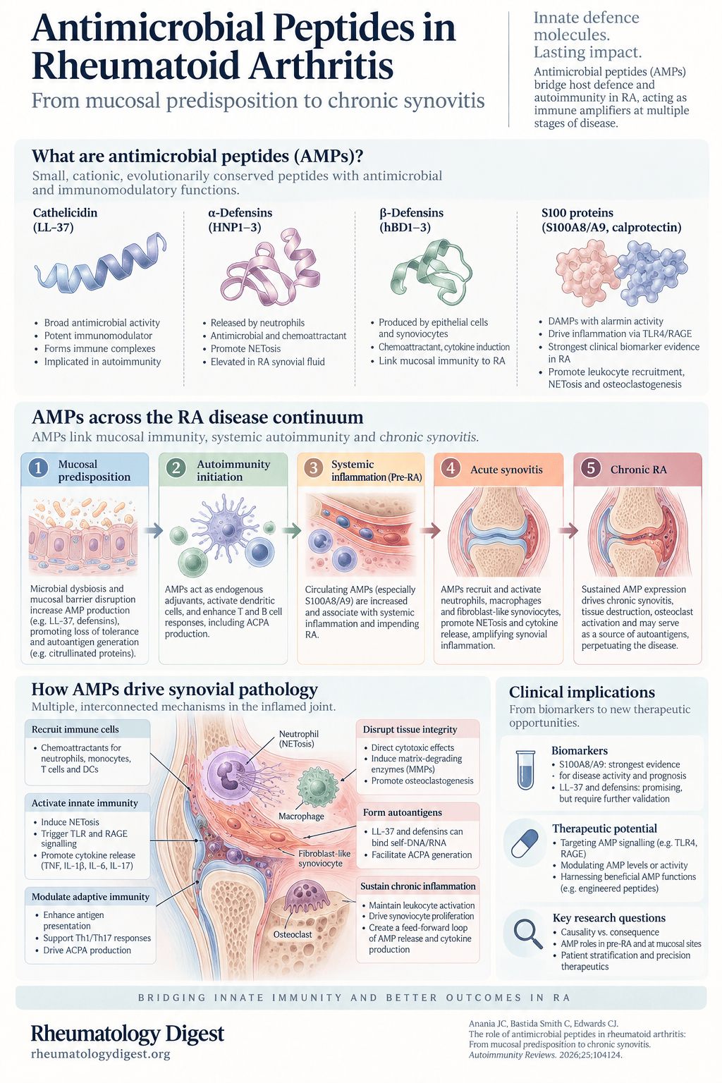
TL;DR: Antimicrobial peptides start as innate defence molecules and end up as autoantigens, immune adjuvants and drivers of bone erosion — but the evidence supports them as amplifiers of RA rather than initiators, and the most immediately usable finding is that calprotectin already beats CRP and ESR for reflecting joint inflammation.
Why these molecules deserve attention
Antimicrobial peptides (AMPs) are small, mostly cationic peptides that constitute a critical arm of innate immunity — direct activity against bacteria, fungi, parasites and viruses, coupled with potent immunomodulation of host cells. They are highly evolutionarily conserved, and their hydrophobic and hydrophilic domains are what let them interact with and disrupt bacterial membranes.
The interesting claim in this review is that the same signals become maladaptive in RA. In the inflamed joint, AMPs disrupt tissue architecture, activate resident and infiltrating immune cells, and — the part that makes them unusual among inflammatory mediators — serve as autoantigens themselves, amplifying the pathogenic immune response they were meant to defend against.
The four families that matter here:
| Family | Members | Principal sources | Upregulated by |
|---|---|---|---|
| Cathelicidin | LL-37 (37 aa, α-helical, net charge +6) | Neutrophils, T cells, B cells, NK cells, eosinophils, adipocytes, keratinocytes, macrophages, mast cells, osteoclasts | Infection, inflammation, vitamin D, ER stress |
| α-defensins | HNP1–3 (29–30 aa) | Neutrophils | Infection, inflammation, tumour microenvironment |
| β-defensins | HBD1–6 (36–50 aa) | Epithelial cells; HBD2 also chondrocytes | Infection via TLR stimulation, TNF, IL-1α, IL-1β |
| S100 proteins | S100A4, A8, A9, A11, A12, B | Monocytes, macrophages, neutrophils, T cells, epithelial cells, osteoclasts | Infection via RAGE/TLR4, inflammation |
S100A8 and S100A9 form the heterodimer familiar in clinic as calprotectin.
One regulatory detail worth noting early: LL-37 is upregulated by vitamin D, but no correlation has been observed between LL-37 levels and serum vitamin D in RA, despite reported associations with vitamin D receptor polymorphisms in some populations. Endoplasmic reticulum stress may be the more relevant inducer — and ER stress is itself associated with RA, promoting AMP production, synoviocyte proliferation and cytokine production.
Where they come from in the joint
Synovial fibroblasts and macrophages are the predominant cell types in the synovium; neutrophils reside mainly in the synovial fluid. Neutrophils are the most prolific AMP source in the RA synovium, and they release AMPs by three distinct routes:
- Primary (azurophilic) granules hold HNP1–3 preformed, released rapidly on degranulation triggered by immune complexes, complement fragments (C5a) and cytokines including TNF and IL-8.
- Secondary (specific) granules contain LL-37. A nice structural detail: granules are released in reverse order to their formation, so secondary granules — formed later, at the myeloblast stage — are more readily secreted than primary granules formed at the promyelocyte stage. Primary granule contents kill and digest pathogens; secondary granule contents are more regulatory.
- NETosis extrudes neutrophil extracellular traps decorated with HNP1–3, LL-37 and citrullinated histones, providing a sustained extracellular AMP depot rather than a bolus.
S100A8/A9 and S100A12 follow a different, non-classical secretory pathway — calcium- and tubulin-dependent, without lysosomal targeting, primed by MAPK signalling (p38, ERK) and reactive oxygen species from the oxidative burst.
Neutrophils from RA patients have a greater propensity to release NETs than healthy controls. And because HNPs and LL-37 both stimulate NETosis as well as being released by it, there is a self-amplifying cycle built into the system from the start.
Two observations argue that neutrophils are not the whole story. Synovial neutrophil counts and HNP1–3 levels do not correlate in RA patients, implying substantial production from other cells. And norepinephrine dose-dependently reduces HNP1–3 secretion in synovial cultures from both RA and osteoarthritis patients, whereas TNF and cortisol reduce HNP1–3 only in OA-derived cultures, with no significant inhibitory effect in RA — suggesting genuinely distinct regulatory mechanisms in degenerative versus autoimmune disease.
The five-stage model
The review’s organising structure is a progression from mucosal predisposition to chronic joint destruction, with AMPs playing a different role at each step. This is the useful part, because it distinguishes when these molecules matter from whether they matter.
1. Pre-clinical predisposition — the mucosal phase
Under physiological conditions LL-37 and HBD-3, produced by synoviocytes and to a lesser extent monocytes and macrophages, maintain barrier integrity and regulate microbial composition.
The hypothesis is that seropositive RA begins outside the joints. In genetically susceptible individuals — HLA-DRB1 shared epitope carriers — smoking and periodontal pathogens induce mucosal stress in lung and oral cavity. ACPAs and rheumatoid factor are detectable in sputum, saliva or serum years before symptomatic RA.
AMPs contribute in two ways here. Certain microbial peptides share structural homology with citrullinated human proteins, supporting molecular mimicry — and pathogen-induced mucosal damage (P. gingivalis orally, P. copri in the gut) can activate autoreactive lymphocytes. AMPs may enhance epitope exposure by disrupting bacterial membranes and increasing antigen availability.
NET formation in RA is strongly dependent on PAD4-mediated chromatin decondensation, and genetic polymorphisms affecting PAD4 expression both increase RA susceptibility and correlate with higher autoantibody titres — linking genetic risk directly to autoantigen generation.
Critically, the immune system is still tolerant at this stage. Tregs function normally. What AMPs do is act as endogenous immune adjuvants: cationic LL-37 and S100 proteins bind extracellular DNA, RNA and citrullinated proteins released from stressed epithelium and NETs, stabilising these autoantigens and promoting their persistence. Acting as opsonin-like bridging molecules, they enhance uptake by antigen-presenting cells via scavenger and Fc receptors. APC function is enhanced — but no sustained adaptive response yet.
The authors characterise this as immune priming and a disturbed immune environment rather than established disease. No one mechanism tips tolerance over into autoimmunity — it is the accumulation of pro-inflammatory signals that leaves the system primed for the next step.
2. Autoimmunity initiation — the break in tolerance
Now innate activation converts antigen exposure into an inflammatory signal.
Self-DNA and RNA released during NETosis, complexed with LL-37, activate APCs through TLR7/8/9, generating a strong type-I interferon response. Notably, DNA complexed with HNPs fails to produce type-I interferon, though it can still activate plasmacytoid DCs — so this is an LL-37-specific property, not a general AMP one.
That interferon signature matters clinically: the peripheral type-I interferon gene signature is detectable years before RA onset and predicts progression to clinical RA. Interferons enhance BAFF production, promote B-cell survival and increase antigen presentation, tying innate nucleic-acid sensing to adaptive autoimmunity.
LL-37 is itself an autoantigen in RA — both IgG and IgM anti-LL-37 autoantibodies are found in patients. It can also be carbamylated in spontaneously generated RA NETs. Carbamylated LL-37 (carLL-37) is internalised by fibroblast-like synoviocytes, loaded into the MHC II compartment, and presented to antigen-specific T cells — activating autoreactive CD4⁺ T cells and correlating with bone erosion. Anti-carLL-37 autoantibodies additionally potentiate osteoclast formation and their capacity to resorb bone.
An isotype detail with mechanistic weight: IgA ACPAs — the predominant mucosal isoform — often precede IgG autoantibodies, implying the initial antigen encounter happens at barrier sites. The transition from IgA to IgG dominance coincides with systemic dissemination and higher disease risk.
3. Systemic inflammation (pre-RA) — innate–adaptive amplification
Systemic immune activation becomes established, but overt synovitis is still limited or absent.
The cytokine effects here are strikingly context-dependent, and the review is careful about it. In macrophages, LL-37 is dose-dependent — moderate concentrations attenuate, high concentrations cause cell damage. It upregulates MCP-1, IL-8, IL-1β, IL-6 and TNF while downregulating MIP1α and IL-12. In monocytes it can raise pro-inflammatory mediators (CXCL1, CCL2, CCL7, IL-6, IL-8) while simultaneously inducing anti-inflammatory IL-10 and IL-19. In IFN-γ-stimulated monocytes it strongly inhibits TNF and IL-12. In mature dendritic cells it suppresses IL-6 and TNF concentration-dependently.
The T-cell picture splits by peptide family, and this is the most interesting divergence in the paper:
- LL-37 induces apoptosis in certain T-cell subsets, including Tregs. Loss or functional impairment of Tregs shifts the balance toward persistent synovial inflammation. LL-37 also skews dendritic cells toward Th17 polarisation, raising IL-17 — which creates a positive feedback loop for further IL-17 and inflammatory cytokine production in cartilage.
- β-defensins do the opposite. HBD-2 and HBD-3 promote T-cell proliferation; HBD-3 enhances IL-2 and also IL-10, possibly a compensatory regulatory signal.
So LL-37 favours loss of regulatory control while β-defensins support expansion and modulation of effector populations — the same broad family pulling in opposite directions.
Neutrophils and B cells sit in close proximity in RA synovial tissue, with direct consequences. Activated synovial neutrophils release BAFF and IL-21, lowering the activation threshold of autoreactive B cells; NETs simultaneously supply citrullinated histones and self-DNA TLR ligands, driving IgM RF and ACPA production. Cathelicidin-deficient mice show markedly reduced IgG1 and IgE responses, implicating AMP-dependent amplification of humoral immunity.
Two downstream consequences worth holding onto:
- ACPA IgG shows reduced galactosylation and sialylation, increasing affinity for Fcγ receptors and complement activation. These glycosylation changes precede disease onset and correlate with severity.
- ACPA immune complexes activate classical complement, generating C3 deposition and C5a-mediated neutrophil recruitment. LL-37 supports C1q in increasing NET-stimulatory activity on macrophages.
An honest gap the authors name: whether AMP-driven class switching arises from direct effects on B cells or indirectly via modulation of APCs and T-cell subsets remains unresolved.
4. Acute RA — synovial damage and clinical onset
Maximal cytokine-driven amplification. The immunopathological focus shifts from mucosal autoimmunity to synovial-centred inflammation, and AMP expression rises sharply within the joint.
The inflammasome is the engine here. Synovial macrophages and fibroblast-like synoviocytes undergo NF-κB-dependent priming in response to AMP-associated cytokines, transcribing pro-IL-1β, pro-IL-18 and NLRP3. DAMPs from damaged joint tissue then trigger NLRP3 assembly and caspase-1 activation, cleaving pro-IL-1β and pro-IL-18 into mature forms — producing the leukocyte recruitment, endothelial activation and matrix-degrading enzyme production behind the rapid swelling and pain of acute synovitis. IL-18 additionally enhances IFNγ and augments T-cell activation. NETs both trigger inflammasome signalling and are further amplified by inflammasome-derived cytokines.
Structural damage follows along several routes:
- IL-6 correlates with increased MMP-13 and cartilage destruction — more collagen II decomposition, less aggrecan.
- TNF is associated with chondrocyte apoptosis, cartilage matrix degradation, less migration of cartilage-derived progenitor cells, and reduced ATP production and mitochondrial membrane potential in chondrocytes.
- S100A8/A9 enhance RANKL expression, linking inflammation directly to osteoclastogenesis and early bone erosion.
- HBD3 activates matrix metalloproteinases to degrade cartilage extracellular matrix.
- LL-37 directly induces apoptosis in osteoblasts — the bone-forming cells — potentially contributing to reduced bone formation in arthritic joints.
HNP levels in synovial fluid are higher in RA patients with erosive disease than non-erosive disease, though no direct effects of HNPs on joint structural cells have been reported.
5. Chronic RA — a self-perpetuating loop
Continuous NET formation ensures ongoing release of autoantigens and DAMPs. The finding I’d single out here: synovial fibroblasts undergo epigenetic reprogramming from chronic activation, and this persists even after inflammatory signals decline. This positional memory is offered as the explanation for why synovitis recurs in previously inflamed joints — a clinical observation most rheumatologists will recognise, now with a mechanism attached.
LL-37 induces osteoblast apoptosis via P2X7 and FPRL1, and triggers secondary neutrophil necrosis, releasing further DAMPs and autoantigens — a destructive positive feedback loop. S100A8/A9 activate macrophages via TLR4, promoting chondrocyte-driven cartilage destruction, while chronic IL-6, TNF and IL-17 exposure stabilises a pathogenic MMP3⁺/IL-6⁺ synovial fibroblast phenotype and suppresses pro-resolving states.
Tertiary lymphoid structures form within the synovial membrane, organised by synovial fibroblasts and follicular dendritic cells. These act as self-perpetuating reservoirs of autoantibody production, maintaining both quantity and quality of the autoantibody response even when systemic inflammation fluctuates. Patients with TLS-positive synovium have high-titre ACPAs and more severe, refractory disease — making TLS a potential therapeutic target in their own right.
The biomarker case, which is the nearest-term application
This is where the review has immediately usable content.
| AMP | Compartment | Finding in RA | Correlation with disease activity |
|---|---|---|---|
| S100A8/A9 (calprotectin) | Serum, synovial fluid, synovial macrophages | Markedly elevated vs healthy controls | Superior to CRP and ESR in reflecting joint inflammation. Correlates with DAS28 (r = 0.50), CRP (r = 0.44), swollen joint count. Independently predicts radiographic joint damage at 10 years. Better predictor of CDAI/SDAI and power Doppler synovitis than CRP |
| S100A12 | Serum, synovial fluid | Elevated vs healthy controls and vs other inflammatory arthritides | Correlates with disease activity, declines with infliximab. Discriminates RA from other inflammatory joint diseases on proteomic profiling |
| S100A4 | Serum | Elevated; expressed by T cells, neutrophils, macrophages | Associated with disease activity; elevated in early RA and may predict insufficient treatment response |
| LL-37 | Synovial tissue, serum/plasma | Elevated in synovial lining and sublining | Anti-carLL-37 titres correlate with bone erosion. Downregulated by TNF inhibitors (adalimumab, etanercept) and intra-articular glucocorticoids — but not by methotrexate. No correlation with serum vitamin D |
| HNP1–3 | Synovial fluid, synovial membrane | High concentrations; higher in erosive than non-erosive disease | No correlation with synovial neutrophil counts |
| HBD1, HBD2, HBD3 | Articular cartilage, synovial membrane | Expressed in RA cartilage; HBD2 induced in chondrocytes by TNF, IL-6, IL-1β | Reflects local cytokine milieu indirectly; no established DAS28/CRP correlation |
Calprotectin is the standout — it is the one AMP here with a genuine claim to clinical utility today, and the 10-year radiographic prediction is the strongest datum in the table.
Therapeutics — the honest position
No AMP-based agent has advanced to a clinical trial in RA since the phase II failure of NI-0101 in 2019. That single sentence is the appropriate calibration for everything that follows.
Preclinical work continues on several fronts:
- Modified LL-37 variants, including citrullination-resistant forms, aiming to retain antimicrobial activity while reducing autoimmune potential.
- LL-37 antagonism — neutralising antibodies to prevent complex formation with DNA/RNA ligands and block downstream TLR7/9 signalling. The authors add a genuinely important caveat: because LL-37 can itself form self-reactive immune complexes, caution is advised when designing antibodies to neutralise it.
- The LL-37-derived peptide IG-19 reduces disease severity and pro-inflammatory cytokines in collagen-induced arthritis.
- Upstream S100 targeting — colchicine effectively prevents S100A8/A9 secretion by inhibiting microtubule polymerisation; neutralising antibodies, peptide-Fc fusions, small-molecule Q compounds and shRNA approaches all exist. But direct blockade of S100 signalling through TLR4/RAGE with neutralising antibodies has not demonstrated clinical benefit.
- Non-human peptides: Scolopendrasin IX, from a centipede used in traditional medicine, binds FPR2 and inhibits joint cytokine production and neutrophil recruitment in murine models — though centipede-derived compounds carry risks of local irritation, allergy and immunogenicity. Rhesus θ-defensin 1 (RTD-1) arrests and reverses joint damage in rodent models while suppressing TNF, IL-1β and IL-6.
Three structural barriers to translation are named plainly: widespread endogenous AMP expression creates off-target and toxicity risk; proteolytic instability is worsened in RA specifically, since patients have elevated MMPs and cathepsins that would degrade therapeutic AMPs — potentially a pathogenic feedback loop; and manufacturing costs remain prohibitive, pushing attention toward computationally designed synthetic analogues.
How much to believe
The authors’ limitations section is unusually candid and worth reading in full. Four points:
- Most mechanistic insight is in vitro or murine — and there is a specific, serious problem here: rodents lack a true LL-37 orthologue. CRAMP is the murine equivalent, but functional divergence between the two peptides means murine models cannot fully recapitulate human LL-37 biology. Given that LL-37 is the most-studied AMP in this field, that constrains a large fraction of the mechanistic evidence.
- Most clinical studies are cross-sectional, precluding causal inference — so it is not established whether altered AMP levels are a driver or a consequence of inflammation. Longitudinal data linking AMP trajectories to onset, progression or treatment response are scarce, and heterogeneity across studies (cohort composition, disease duration, concomitant therapy, disease activity at sampling, and assay methodology — ELISA vs mass spectrometry vs immunohistochemistry vs gene expression) limits comparability and complicates meta-analysis.
- Most cohorts do not stratify by sex, menopausal status or ethnicity, and published data cluster around a handful of well-characterised peptides (LL-37, HNP1–3, S100A8/A9) while the broader defensin and S100 families remain substantially understudied.
- The regulatory complexity is the deepest problem. These peptides act as both pro- and anti-inflammatory mediators depending on concentration, cell type and microenvironment — from which the authors draw the uncomfortable conclusion that where AMP regulation goes wrong in an individual patient, the overall immunological result may not be predictable at all.
That last point deserves emphasis, because it cuts against therapeutic targeting more than any of the individual negative trials do. A molecule that suppresses IL-6 and TNF in mature dendritic cells while upregulating them in macrophages is not a straightforward drug target.
What to take from this
- The framing claim is that AMPs are amplifiers, not initiators. The review states plainly that the evidence to date casts these peptides as escalators and modulators of an immune response rather than the thing that starts the disease on its own. Read the mechanistic sections with that calibration.
- Calprotectin is the practical takeaway. Superior to CRP and ESR for reflecting joint inflammation, correlates with DAS28 and swollen joint count, and independently predicts radiographic damage at 10 years. This is the one item here that could change a monitoring decision now.
- LL-37 responds to TNF inhibitors and intra-articular steroids but not methotrexate — a small observation, but one that implies AMP suppression is not a generic consequence of disease control.
- The mucosal-origins story gains mechanistic detail. IgA ACPAs preceding IgG, molecular mimicry between microbial peptides and citrullinated human proteins, and PAD4-dependent NETosis linking genetic risk to autoantigen generation all fit the same account of RA beginning at barrier surfaces — the same argument made from the pulmonary side in lung disease in RA.
- Epigenetic reprogramming of synovial fibroblasts offers a mechanism for why synovitis recurs in previously affected joints, which is worth knowing even though nothing follows from it therapeutically yet.
- The open questions the authors name are the right ones: which AMPs are viable therapeutic targets versus biomarkers only; whether AMP profiles predict response to anti-TNF, anti-IL-6 or JAK inhibitors (unexplored in prospective cohorts); and sex-specific differences in AMP expression, noted in other autoimmune diseases but never systematically evaluated in RA despite its strong female predominance — which connects directly to the gap identified in sex-dependent mechanisms in rheumatic disease.