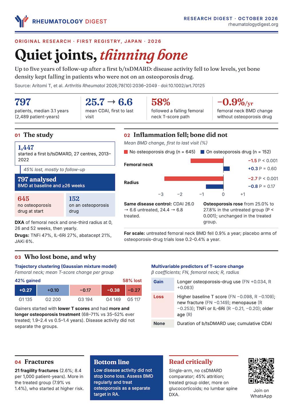
TL;DR: In a Japanese RA cohort, bone density kept falling over about three years on a biologic or targeted DMARD even though disease activity became low — and only patients also on osteoporosis treatment held their bone.
The reasoning that inflammation control should protect bone is sensible, and several small studies supported it. This group’s earlier one-year report did not, and this paper extends the same cohort to as long as five years. The result is a reminder that osteoporosis in RA has more than one cause, and that a good CDAI is not a bone outcome.
The question and the earlier work
Systemic osteoporosis is common in RA, with a prevalence roughly twice that of the general population, and it causes fractures that add to the disease burden and reduce physical function. Contributors include age, menopause, smoking, low physical activity, glucocorticoids, chronic kidney disease, diabetes and chronic inflammation. Treatment rates remain low; even among glucocorticoid users, only 38–60% receive osteoporosis therapy.
Because inflammation is one cause of systemic bone loss, it was hypothesised that suppressing it with biologic or targeted synthetic DMARDs (b/tsDMARDs) would also benefit bone, and several small studies found that bone density stabilised or improved on these drugs. Against that, recent fracture studies show that although b/tsDMARD use has increased and remission rates have roughly doubled, fracture rates have also risen. The group’s previous report in 1,164 patients treated for one year without antiosteoporotic drugs found that BMD of the femoral neck and radius fell significantly despite a marked reduction in disease activity. This study asks whether that continues over longer follow-up.
Design
This was a multicentre, prospective observational study within the FIRST registry, an extension of the earlier work, run through the University of Occupational and Environmental Health in Kitakyushu and 27 institutions within 150 km. Patients with active RA by the 2010 criteria who were starting their first b/tsDMARD between August 2013 and July 2022 were enrolled consecutively. The drugs were TNF inhibitors (infliximab, etanercept, adalimumab, golimumab, certolizumab, ozoralizumab), IL-6 receptor inhibitors (tocilizumab, sarilumab), abatacept, and JAK inhibitors (tofacitinib, baricitinib, peficitinib, upadacitinib, filgotinib). There were no restrictions on other treatments, including osteoporosis drugs.
BMD was measured by DXA at the femoral neck and one-third radius at baseline, weeks 26 and 52, then annually for up to five years. Lumbar spine was deliberately not measured, to avoid overestimation from degenerative disease and vertebral compression fractures, and bone turnover markers were not evaluated. The primary endpoint was the change in T score; secondary endpoints included Z score, percentage change in BMD, the proportion changing beyond the least significant change (LSC), and factors associated with T score change. Statistics included inverse probability of treatment weighting, multivariable regression, and a Gaussian mixture trajectory analysis, with false discovery rate correction for multiple comparisons. The treatment-group comparison was by use of osteoporosis medication at enrolment.
Who was analysed
Of 1,447 enrolled patients (all Asian), 797 were analysed, those with BMD at baseline and at least once at 26 weeks or later. About 45% of the enrolled patients dropped out, most commonly by loss to follow-up; the authors report limited differences between enrolled and analysed patients and assume random censoring.
- Mean age 61.4 years, 77.2% women, mean disease duration 60.9 months, mean CDAI 25.7 at baseline (high disease activity)
- 74.2% on methotrexate (mean 12.4 mg/week), 22.0% on glucocorticoids (mean prednisolone-equivalent 9.1 mg/day)
- Baseline osteoporosis in 31.7%, a history of fracture in 18.1%, 70.9% of the women postmenopausal
- b/tsDMARDs: TNF inhibitors 373, IL-6R inhibitors 211, abatacept 165, JAK inhibitors 48
- Median follow-up 3.1 years (2,489 patient-years); patients were on a b/tsDMARD for 83.3% of the observation period
Two groups by osteoporosis treatment at baseline: 645 patients not on osteoporosis treatment (antiosteoporosis−) and 152 on it (antiosteoporosis+). The treated group was older (69.8 vs 59.4 years), more often women (90.1% vs 74.1%), with longer disease (98.0 vs 52.1 months), more advanced stage, more glucocorticoid use (52.0% vs 14.9%), more menopause and fracture history (40.8% vs 12.7%), and much lower baseline T scores (femoral neck −2.22 vs −1.43). Treatments in the treated group were bisphosphonates (72.4%), active vitamin D (38.8%), denosumab (11.2%), with smaller numbers on selective estrogen receptor modulators and parathyroid hormone. Some patients took more than one.
Disease activity improved; bone did not
CDAI fell from 25.7 to 6.6 (P < 0.001) over the observation period, with a mean CDAI during observation of 8.2. In the whole analysed group:
- Femoral neck T score fell from −1.58 to −1.67 (P < 0.001); BMD change −1.2%, or −0.7% per year
- Radius T score fell from −0.96 to −1.25 (P < 0.001); BMD change −2.3%, or −1.0% per year
- Total hip T score also fell, from −1.14 to −1.21
- The prevalence of osteoporosis rose from 31.7% to 34.6% (P < 0.001)
- 21 new fragility fractures (2.6%) occurred: 12 vertebral, 1 distal radius, 8 hip
58% of patients (460) had a decrease in T score overall.
The split that matters: treated versus untreated
| Antiosteoporosis− (n = 645) | Antiosteoporosis+ (n = 152) | |
|---|---|---|
| CDAI | 26.0 → 6.6 | 24.4 → 6.8 |
| Femoral neck T score | −1.43 → −1.54 (P < 0.001) | −2.22 → −2.21 (P = 0.830) |
| Radius T score | −0.60 → −0.93 (P < 0.001) | −2.49 → −2.59 (P = 0.071) |
| Femoral neck BMD change | −1.5% | +0.3% (NS) |
| Mean duration of osteoporosis treatment | — | 3.1 years |
Both groups reached low disease activity, but bone loss was confined to the untreated group. In the treated group, the yearly BMD change at the femoral neck was 0.0%. In the untreated group it was −0.9% per year, which the authors note is faster than the placebo rates in osteoporosis drug trials (−0.2% to −0.4% per year). Osteoporosis prevalence rose from 25.0% to 27.8% in the untreated group but did not change significantly in the treated group (60.5% to 63.8%).
Z scores tell the same story. The Z score compares a patient with the average for their age and sex, so a falling Z score means loss beyond ageing. It fell at the radius in the untreated group (0.91 to 0.81), whereas it rose at both sites in the treated group. In the untreated group the femoral neck Z score rose slightly (−0.23 to −0.18) even as the T score fell, so the femoral neck result is mixed.
Beyond measurement error. Using the least significant change, BMD in the whole group at the femoral neck increased in 14.2%, was stable in 58.8% and decreased in 27.0%; at the radius, 18.6%, 42.4% and 39.0%. In the treated group the proportion decreasing was lower (femoral neck 20.4% against 28.5% untreated) and the proportion increasing was higher (19.1% against 13.0%).
Confounding was addressed, to a point. After inverse probability weighting on propensity scores, the favourable bone outcomes in the treated group persisted, and the weighting achieved adequate balance on the measured covariates.
What predicted a change in T score
In multivariable analysis:
- Longer duration of osteoporosis treatment was associated with a higher T score at both the femoral neck and radius.
- A higher baseline T score was associated with a subsequent decrease at both sites.
- At the radius, older age, menopause, and TNF or IL-6 receptor inhibitor use were associated with a decrease. At the femoral neck, a new fracture during follow-up was associated with a decrease.
- Duration of b/tsDMARD use did not predict bone maintenance, and neither did mean or cumulative CDAI.
The authors also ran a trajectory analysis (Gaussian mixture modelling), which grouped femoral neck BMD into five patterns and radius into four. Groups whose T score increased had lower baseline T scores, more osteoporosis medication at baseline and throughout, and longer treatment durations; groups whose T scores fell were characterised by the reverse. In the femoral neck, the groups that increased (about 42% of patients) had a mean osteoporosis drug use of 1.9 and 2.4 years; the decreasing groups 1.4, 0.7 and 0.5 years. Average CDAI during observation was low in the first four femoral neck groups (about 7–8); the fifth, which lost bone and had the shortest follow-up (1.4 years), averaged 12.0 with higher CRP and HAQ, but in the multivariable analysis cumulative CDAI was not associated with T score change.
Patients with new fractures were older, more often women and postmenopausal, more often with reduced eGFR, more often on glucocorticoids (38.1% vs 21.5%) and more often with a prior fracture (61.9% vs 16.9%), with lower baseline T scores. There was no obvious difference in fracture sites between treated and untreated patients. The fracture incidence was 8.4 per 1,000 person-years, below the 13.0–21.0 per 1,000 reported in earlier RA studies.
How to read it
What the data support. In patients with RA on a first b/tsDMARD, whose disease activity became low, a majority (58%) lost bone density at one or both sites, and bone was maintained mainly in patients who were also receiving osteoporosis treatment. Neither how long they stayed on the b/tsDMARD nor how well their disease was controlled predicted bone outcome.
Three cautions the authors state, and one more worth adding:
- There was no comparison group on other treatment, such as csDMARDs. The design is single-arm and observational, so it cannot establish whether b/tsDMARDs help, harm or do nothing for bone. The title’s claim that these drugs “do not arrest” bone loss is about what happened on them, not a comparison with anything.
- The treated and untreated groups differed substantially. The treated group was older and at much higher fracture risk, and treatment was not randomised. Medication use was unrestricted, so some untreated patients began osteoporosis drugs during follow-up (the rate rose from 0 to 45.6%), which could blur the contrast; the authors judge this unlikely to change the conclusion.
- Fractures were few and self-reported. Twenty-one events, from patient reports and physician records, cannot support conclusions on fracture outcomes. Asymptomatic fractures were not captured.
- Attrition was high. Only 797 of 1,447 patients (55%) were analysed, mostly because follow-up BMD was missing. The authors assume the missingness was random, which cannot be confirmed.
- Other limits: all patients were Japanese, and the authors attribute the difference from a Taiwanese study that did show maintained BMD to differences in age, smoking and osteoporosis treatment (dietary calcium intake was comparable); the relation between BMD change and fracture is unclear in this cohort, because the untreated group lost more bone but had fewer fractures than the treated group (who had higher baseline risk); and dose and adherence data for osteoporosis drugs were lacking.
One feature of the findings to keep in view: the untreated group’s bone loss was modest in absolute terms, about 1.5% at the femoral neck over the observation period and under 1% per year, and the authors say plainly that its clinical impact on fractures remains unclear.
What it means for practice
The authors’ conclusion is that osteoporosis should be treated as an independent aspect of RA management, beyond inflammation control: BMD declined in patients not on osteoporosis medication even after b/tsDMARDs brought disease activity down, while concomitant osteoporosis treatment helped maintain bone. They support regular BMD assessment, noting that DXA involves low radiation, and propose every one to two years for high-risk patients.
The reasonable reading is practical rather than mechanistic. A patient with RA whose CDAI has come down on a biologic or JAK inhibitor should not be assumed to have protected bone, and the decision to measure and treat bone should rest on the usual fracture-risk factors: age, menopause, prior fracture, glucocorticoid exposure and low baseline BMD. What the study cannot say is whether any particular b/tsDMARD class is better or worse for bone, whether treating the patients who were left untreated would have prevented fractures, or how much of the loss was inflammation, ageing or other causes.
Disclosures noted in the source: support from a grant-in-aid for scientific research and from the University of Occupational and Environmental Health, Japan; author disclosures are available online with the paper.