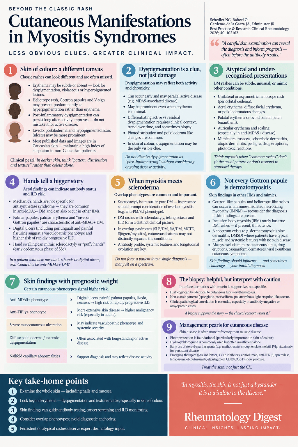
TL;DR: A careful skin examination can identify life-threatening myositis phenotypes weeks before antibody testing returns — and can be right when the antibody panel is falsely negative.
The pattern of muscle involvement and the autoantibody profile are how inflammatory myopathies are usually sorted. This review argues that a thorough examination of the whole integumentary system — scalp to toe, including nails and oral mucosa — does work the antibody panel cannot, because it is available immediately and the panel is not. In anti-MDA5+ disease with rapidly progressive ILD, that gap is measured in weeks and the stakes are transplant evaluation.
The skin biopsy helps, but less than you would expect
A skin biopsy should be considered whenever a patient with inflammatory myopathy has suggestive skin findings. The classic reaction pattern in dermatomyositis is vacuolar interface dermatitis with extracellular mucin deposition, and a concordant biopsy supports the diagnosis — particularly in antibody-negative, amyopathic or clinically equivocal cases.
The problem is specificity in both directions.
The vacuolar interface pattern is not specific to DM. It is seen in cutaneous lupus erythematosus, graft-versus-host disease, viral exanthems, drug eruptions, cutaneous lymphoma (mycosis fungoides) and erythema multiforme. Mucin deposition is not specific to DM or CLE either, and appears in eczematous rashes and photodamaged skin. Light microscopy findings in DM can be identical to those in CLE — and the interface dermatitis pattern was observed in 82% of patients in a DM cohort who had originally been misdiagnosed, mostly as CLE or undifferentiated connective tissue disease.
There is a counter-intuitive consequence the authors highlight: skin biopsies with features consistent with dermatomyositis are associated with longer diagnostic delays than inconsistent biopsies. The likely explanation is that a biopsy compatible with lupus anchors the clinician on lupus in a patient who actually has DM. When seeing a patient referred for presumed CLE, the review’s advice is to resist anchoring bias and diagnostic momentum by explicitly considering unrecognised DM.
Running the other way, biopsies from unequivocal DM can fail to show interface dermatitis at all, instead resembling polymorphous light eruption or other reaction patterns (spongiotic, psoriasiform). Hence the practical rule: a patient with biochemical or clinical evidence of myositis plus a classic dermatomyositis rash can be confidently diagnosed with DM, with or without supportive skin histopathology.
The pathognomonic rashes
DM is the prototype IIM with skin involvement, which may be easily recognisable, very subtle, or absent entirely — “dermatomyositis sine dermatitis”. The findings sort into pathognomonic and characteristic groups.
Gottron’s papules are erythematous to violaceous flat-topped papules over the extensor surfaces of the MCP and interphalangeal joints. The distribution is a useful discriminator: Gottron’s papules sit over the joints, whereas CLE tends to involve interphalangeal or interarticular skin with relative sparing over the MCP/PIP joints. Gottron’s sign describes erythematous to violaceous patches and macules over extensor joint surfaces such as elbows and knees, and is sometimes mistaken for plaque psoriasis.
The heliotrope rash is violaceous erythema of the upper eyelids, named for the purplish flowers of Heliotropium arborescens. There may be periorbital oedema, and the rash can extend to the forehead, inferior eyelids, medial cheeks and nose. In skin of colour the erythema may appear subtle or dusky. The atypical variants are worth knowing because they are easy to dismiss: a completely unilateral heliotrope, bilateral periorbital oedema without any rash, and unilateral periorbital oedema. Oedematous involvement of the perinasal area, mid-face and lips can also occur with or without periorbital involvement, mimicking angioedema. These subtle or unusual variations can be the sole initial manifestation of DM.
The characteristic rashes
Photodistributed erythema and poikiloderma produce the “V sign” of the anterior neck and upper chest and the “shawl sign” of the posterior neck, shoulders and upper back. These need distinguishing from poikiloderma of Civatte — chronic photodamage, which lacks pruritus and acute onset and comes without other DM features. The halter sign, or reflected shawl sign, is the classic shawl sign with sparing of the mid-back and then diffuse involvement of the lower back. A symmetric poikilodermatous to papular rash of the lateral thighs is the holster sign.
Nailfold involvement is common — ragged cuticles, haemorrhagic nailfold infarcts, prominent periungual erythema and telangiectasia — though these gross findings also occur in systemic sclerosis and SLE. The high prevalence of a scleroderma pattern on nailfold capillaroscopy in DM helps differentiate it from polymyositis, and certain capillary alterations over time allow some separation from SSc. Capillaroscopic findings in DM tend to improve with overall disease control.
The scalp is commonly affected and frequently mimics psoriasis, seborrhoeic dermatitis, alopecia and other inflammatory scalp conditions. It can present as diffuse psoriasiform plaques, or as intensely pruritic and often treatment-resistant poikiloderma and telangiectasia of the scalp.
Three findings that carry more weight than they get
Pruritus in DM is prominent, correlates with disease severity, and produces a lower quality of life than pruritus from atopic dermatitis or psoriasis. Compared with SLE, patients with DM have more prevalent, more severe and more treatment-resistant pruritus — which is genuinely useful when trying to separate amyopathic DM from CLE. One caution: angulated excoriations should not be mistaken for spontaneous vasculopathic ulceration, since a history of scratching can usually be elicited.
Poikiloderma is the triad of epidermal atrophy, dyspigmentation (both hyper- and hypopigmentation) and telangiectasia. It may occur alongside papular changes at typical DM sites — chest, neck, upper back, thighs — or appear more extensively elsewhere.
Calcinosis cutis is abnormal deposition of insoluble calcium salts in skin, subcutaneous tissue, tendons, fascia and muscle. It is most common in inadequately controlled DM but also occurs in antisynthetase syndrome, SSc and MCTD. It presents as superficial papules and nodules, deep subcutaneous lesions, or diffuse myofascial plane deposits, and calcifications may extrude through the skin with consequent infection and ulceration.
Rarer findings include “whip-like” flagellate erythema, lobular panniculitis, oral mucosal ulcers, erythroderma, and follicular keratotic papules mimicking pityriasis rubra pilaris (“Wong-type DM”).
What else it might be
Cutaneous DM is often misdiagnosed as CLE, and may be labelled UCTD if a myositis-specific workup is not done. Anti-TNF therapy can induce DM, psoriasiform eruptions and drug-induced lupus, all of which closely mimic each other. Other mimickers include plaque psoriasis, atopic dermatitis, rosacea, pityriasis rubra pilaris, airborne or allergic contact dermatitis, acanthosis nigricans, phototoxic drug eruption, polymorphous light eruption and poikilodermatous mycosis fungoides.
Two systemic mimickers are worth carrying. Pellagra — niacin deficiency — produces a symmetric bilateral photodistributed rash over the upper chest and neck resembling the shawl and V signs. Trichinosis produces periorbital and facial oedema alongside myalgia, CK elevation, conjunctivitis, fever and rash. Mimickers of Gottron’s papules include verrucae, knuckle pads, erythema elevatum diutinum and multicentric reticulohistiocytosis, while severe mechanic’s hands and hiker’s feet can mimic an acquired palmoplantar keratoderma.
Anti-MDA5+ DM: recognise the phenotype, not the antibody
This is the section that changes practice.
Early visual recognition of anti-MDA5+ DM as a phenotypic syndrome rather than an antibody result is critical given the risk of rapidly progressive ILD. The reasons are logistical as much as clinical:
- Anti-MDA5 antibodies fluctuate, can occur in low titres, and fail to be detected by line blot assays in 17% of cases. Negative testing against highly suggestive clinical features should prompt repeat testing.
- Turnaround time ranges from two to eight weeks — an unacceptable delay when the competing priorities are disease stabilisation and early transplant evaluation.
The extracutaneous profile includes fever, lymphopenia, elevated ESR, elevated liver enzymes, hyperferritinaemia, hand swelling with peripheral and acral arthritis, dyspnoea at presentation, ILD — and, notably, a lower prevalence of myositis.
The cutaneous signs that occur more frequently in anti-MDA5+ than anti-MDA5− DM are: mechanic’s hands, palmar papules, palmar erythema, diffuse alopecia, oral mucosal pain and ulceration, and vasculopathic ulcerations overlying Gottron papules, the elbow, or the digital pulp and periungual region. Critically, the pathognomonic signs — Gottron’s papules/sign and heliotrope — occur in both groups with no statistically significant difference in prevalence. They do not help you here; the findings above do.
Several deserve individual attention:
- Hand swelling is significantly associated with anti-MDA5+ DM and can mimic the “puffy hands” of early oedematous SSc.
- Ulceration distributed over the Gottron papules of the dorsal hands is particularly recognisable and characteristic, though ulcers also occur with malignancy and other antibody profiles. Ulceration and livedoid lesions similarly affect periungual skin, digital pulp, feet and toes. The presence of these vasculopathic ulcers is associated with increased risk of ILD.
- Mechanic’s hands occur more often in anti-MDA5+ DM than other DM subtypes, but because they also appear in antisynthetase and scleromyositis syndromes, the review’s framing is that they should be read as a high-risk finding for underlying ILD independent of autoantibody status.
- The palmar surface deserves specific inspection. Painful erythematous papules are common there; over the flexural interphalangeal joints they are termed “inverse Gottron papules” and may be hyperkeratotic or ulcerative. Papules can also appear on the lateral digit. These palmar and lateral digital findings have been described in MDA5-associated rapidly progressive ILD even without pathognomonic rashes such as the heliotrope or Gottron’s sign. Palmar erythema is an independent predictive factor for acute/subacute ILD and correlates with anti-MDA5 positivity.
- Oral involvement includes gingival telangiectasia, ulceration and oral pain, with ulcers more frequent than in other DM cohorts.
- Auricular involvement is newly described: ten reported cases of erythematous papules with or without ulceration favouring the helix and antihelix. Across those ten, ILD was universal and half (5/10) died of rapidly progressive ILD.
Malignancy, TIF1γ and NXP2
The cutaneous signs more prevalent in paraneoplastic DM are poikiloderma, periungual erythema, cutaneous ulceration, rapid onset of symptoms, and typical skin involvement (Gottron’s sign, centrofacial erythema, erythema of the arms and forearms). A useful distinction: the ulcerations in malignancy occur on skin surfaces other than the typical anti-MDA5+ sites of hands and extensor surfaces. Conversely, higher prevalence of pruritus, clinically amyopathic DM and oral manifestations correlates with DM without malignancy.
Anti-TIF1γ and anti-NXP2 are the antibodies most frequently detected in paraneoplastic DM. Findings correlating with anti-TIF1γ+ DM include diffuse photoerythema, psoriasiform lesions, scalp rash, facial rash, V-neck sign, back rash, the holster sign, and a characteristic asymptomatic poikilodermatous variation termed “red on white” patches. Verrucous, non-tender palmar lesions can occur — in direct contrast to the painful erythematous palmar papules of anti-MDA5+ DM, and palmar erythema negatively correlates with anti-TIF1γ positivity. The distinctive finding is the “ovoid palatal patch”, a violaceous ovoid patch of the hard palate; recognising it should raise concern for TIF1γ and its association with paraneoplastic disease.
Anti-NXP2+ DM carries a higher prevalence of calcinosis cutis and subcutaneous oedema that may be generalised or favour the limbs, mimicking deep vein thrombosis. It may also present as a polymyositis phenotype with or without facial oedema and periungual erythema.
Skin of colour
Dyspigmentation (dyschromia) is a postinflammatory mixture of hyper- and hypopigmentation, more pronounced in patients with skin of colour. It arises from chronic interface dermatitis affecting melanocytes and melanosome-containing keratinocytes, causing pigment incontinence into the dermis.
In case series and reviews of DM in patients with skin of colour, dyspigmentation was a frequent feature alongside classic findings like the shawl sign, V sign and facial erythema. Two points follow. Marked dyspigmentation may be the key clinical finding when violaceous erythema is less evident. And dyspigmentation should not be read as inactive damage — it can occur with ongoing myositis and cutaneous inflammation.
Two signs were identified in a retrospective study of DM patients of Hispanic ancestry with skin of colour. The sunburn sign is bright red erythema on the forehead, cheeks, nose and chin; the suntan sign is its likely sequela, brown-grey hyperpigmentation with mild desquamation in the same areas. Of 37 patients evaluated, sunburn sign was seen in 18 and suntan sign in 22. Both may aid diagnosis in patients in whom a typical DM rash can simply appear as tanned skin.
Juvenile DM exhibits the same pathognomonic, characteristic and nonspecific rashes seen in adults. Severity of skin damage was notably higher in an adult cohort than in JDM (70.3% versus 43.9%), reflected in increased cutaneous scarring and poikiloderma. Although severe calcinosis is seen in uncontrolled JDM, one cohort study found no statistically significant difference in calcinosis between JDM and adult DM.
Antisynthetase syndrome: mechanic’s hands are not the hallmark
Antisynthetase syndrome is distinguished by antibodies against cytoplasmic tRNA synthetases, fever, myositis, inflammatory arthritis, Raynaud phenomenon and ILD. Its most widely recognised cutaneous finding is mechanic’s hands — hyperkeratotic fissured plaques along the lateral aspect of the digits, with the equivalent on the feet termed mechanic’s feet or hiker’s feet. Vasculopathic and periungual findings occur too: periungual erythema, capillary loop distortion and haemorrhage, with digital ischaemic changes reported in association with severe systemic disease.
But the conventional teaching does not survive the cohort data. Approximately one-third of ASyS patients had DM-like cutaneous involvement in a retrospective analysis, some of it DM-specific. In a pooled cohort of 1,462 patients with antisynthetase antibody positivity, only 28% exhibited mechanic’s hands compared with 32% who had typical cutaneous manifestations of DM. In another group of 132 ASyS patients, 62% had DM-specific skin involvement against 58% with mechanic’s hands. Multiple cohorts therefore show DM-specific rashes occurring at higher or similar frequency to mechanic’s hands in ASyS — which directly challenges the notion that mechanic’s hands are the hallmark cutaneous sign.
This matters beyond taxonomy. In the pivotal phase 3 ProDERM trial supporting the FDA-approved DM indication for 10% IVIg, exclusion criteria included polymyositis and overlap myositis — yet every patient exhibited a “typical skin rash”, and at least 11 enrolled DM patients had an antisynthetase antibody as their only myositis-specific antibody. Whether such patients are “lumped” into DM or “split” into a non-DM overlap spectrum has real consequences for clinical trial eligibility and insurance coverage.
Overlap myositis, and the value of sclerodactyly
Systemic sclerosis. Myositis occurs in 9% to 17% of large SSc cohorts, negatively correlated with anticentromere antibodies and positively associated with U1RNP, U3RNP, PM/Scl, RuvBL1/2 and Ku. Diffuse cutaneous disease occurred in 60% of SSc-associated myopathy versus 32.6% without myopathy.
Sclerodactyly is the discriminating sign. In a meta-analysis of 3,487 patients it was found in 0% of anti-Mi2 and only 12% of antisynthetase patients, versus 76% in anti-PM-Scl, 59% in anti-U1RNP and 38% in anti-Ku. Since fibrotic skin disease is not generally observed in pure DM (<2%), the presence of sclerodactyly should alert the clinician to an overlap myositis syndrome.
The anti-PM/Scl syndrome is a distinct phenotype: only 30% met full SSc criteria, but 66% featured sclerodactyly, 85% had DM-specific skin findings, and 80% had mechanic’s hands — higher than the 58% in a comparator ASyS group. It matters because of the tendency to ILD (61%) and significant muscle weakness (93%) with a unique pattern in which arm abductors are weaker than hip flexors. In a group of 41 such patients, at least 21 (51%) had sclerodactyly plus either a heliotrope or Gottron rash, validating a sclerodermatomyositis phenotype.
Isolated anti-Ku+ SSc-like disease features frequent sclerodactyly, limited cutaneous SSc, myositis and ILD, without DM-specific rashes. Notably, digital ulcers are much less prevalent in SSc-like conditions with overlap myositis — 5% in anti-PM/Scl and 7.7% in isolated anti-Ku+, against 55% in a conventional anticentromere-positive SSc group.
MCTD. Lifetime prevalence of myositis approaches 51%, possibly as high as 77% in certain subgroups. In 79 patients meeting Kasukawa’s criteria, CLE-like lesions were seen in 54.4%, SSc-like in 60.8%, and DM-like in only 5% — so MCTD presents lupus-like and SSc-like skin manifestations far more often than DM-like rash.
SLE. Myositis prevalence in a retrospective review of 1,718 SLE patients was 6.3%, with 47% having CLE, 41% SSc-associated cutaneous manifestations, and 31% dermatomyositis-specific rashes — notable enough that 33 patients meeting SLICC criteria also exhibited DM-specific rashes. Two cautions: findings interpreted as SLE- or CLE-specific also occur in anti-MDA5+ DM (circular scarring lesions mimicking discoid CLE), which limits their applicability to SLE classification; and weakly positive anti-dsDNA antibodies are known to occur rarely in anti-MDA5+ DM, so serologies alone should be interpreted with caution. SLE-specific organ damage is a more compelling basis for genuine overlap.
Rheumatoid arthritis. IIM prevalence in a cohort of 350 RA patients was 1.4%. The most relevant overlap occurs in anti-MDA5+ DM, where anti-CCP positivity portends a higher risk of arthritis that is often the first symptom — and, importantly, dual anti-MDA5+/anti-CCP+ positivity carries a higher malignancy risk than isolated anti-MDA5+ DM, which is not typically associated with malignancy. DM/RA overlap needs differentiating from anti-TNF drug-induced DM, which typically requires drug discontinuation.
Sjögren’s syndrome. The relationship is variable — subclinical myositis reported in up to 72% in some studies, while another 5-year cohort found definitive myositis in only 1% of primary SjS. Annular erythema of Sjögren’s syndrome has been reported to correlate with myositis activity in one patient, though whether it is distinct from CLE remains controversial.
The myopathies where skin should make you think again
Polymyositis is characterised by lack of skin involvement. Its notable mimicker is dermatomyositis sine dermatitis (DMSD) — associated with anti-NXP2, lacking the pathognomonic rashes, but showing DM-specific muscle biopsy findings including perimysial atrophy with or without perimysial perivascular infiltration. Even so, about 70% of anti-NXP2+ DMSD patients still exhibit some skin or subcutis involvement (facial oedema, periungual erythema, subcutaneous oedema), and some develop skin involvement later — 0.5 to 32 months after muscle biopsy diagnosis. The authors suggest some DMSD may represent a forme fruste of skin involvement, analogous to the subtle myositis of hypomyopathic DM.
Immune-mediated necrotising myopathy is defined by muscle fibre necrosis with minimal inflammation, associated with anti-HMGCR or anti-SRP. Extramuscular involvement is rare and mild if present at all: in two retrospective cohorts of 115 and 135 antibody-positive subjects, heliotrope or Gottron’s rashes were seen in approximately 5% and 4% respectively. Non-specific rashes are rarely described and may simply represent concurrent unrelated skin disease.
Inclusion body myositis features slowly progressive, typically asymmetric weakness of the proximal quadriceps and distal finger flexors without skin involvement. The rule the authors state plainly: if skin findings appear in a patient suspected of having IBM, consider an alternative diagnosis — dermatomyositis, overlap myositis, or a rash unrelated to IIM.
Managing the skin
Skin involvement may precede myositis, develop alongside it, or occur without it in amyopathic DM, and in classic DM with myopathy the skin can improve in parallel with the muscle. But many patients have a refractory cutaneous course: in a 3-year follow-up of 74 DM patients, only 38% experienced clinical remission of cutaneous disease. Factors associated with achieving remission were DM-associated malignancy, increased age, and mycophenolate mofetil at 3 g/day; anti-MDA5+ patients had lower remission rates regardless of vasculopathic ulceration.
The goals are avoiding flares, preventing irreversible damage (dyspigmentation, scarring, calcinosis), and relieving pruritus, pain and the psychosocial burden of visible disease. When cutaneous disease is severely refractory, the review suggests checking three drivers before escalating: nonadherence to medication, nonadherence to photoprotection, or an undetected or inadequately controlled myopathy requiring systemic corticosteroids.
DM is an exquisitely photosensitive disease, and strict photoprotection is a cornerstone of management — broad-spectrum sunscreen, photoprotective clothing, and sun avoidance. Patients should also avoid non-solar ultraviolet sources including tanning beds, nail lamps and older photocopy devices.
Established therapies. Topical corticosteroids and calcineurin inhibitors are frequently prescribed but rarely used in isolation except in mild skin-limited disease, and adherence to topicals is low even in severe inflammatory skin disease. Systemic corticosteroids are warranted for myopathy or ILD but are generally avoided as skin-directed therapy given long-term safety and limited efficacy in refractory cutaneous disease without active myositis.
Hydroxychloroquine is a common first-line systemic therapy for amyopathic DM without ILD, but monotherapy is often inadequate — upward of 88% of cases require a second agent. It is further complicated by a considerable 31% prevalence of HCQ-induced cutaneous reaction distinct from the existing DM rash, an adverse effect that appears uncommon among patients treated with HCQ for CLE.
Steroid-sparing agents include methotrexate, azathioprine and mycophenolate mofetil, with mycophenolate at 3 g/day particularly effective in refractory cutaneous disease. Methotrexate has historically been used cautiously with ILD, but a recent study of 163 DM patients reported no significant difference in ILD risk versus other immunosuppressants. Oral sirolimus has been described in a small case series. Tacrolimus and cyclophosphamide are typically reserved for amyopathic cases with severe ILD.
Rituximab showed skin improvement in 81% of patients in a meta-analysis of 26 studies, against 59% and 65% improvement in muscle and lung disease respectively. IVIg works best where baseline cutaneous activity is higher: 87% of patients started on IVIg for refractory cutaneous disease improved after a mean of 1.82 ± 1.38 cycles.
The vasculopathic ulcerations of anti-MDA5+ DM may be refractory, with adjuncts including intralesional triamcinolone, botulinum toxin, and vasodilators (nifedipine, sildenafil, intravenous prostaglandins, bosentan). Calcinosis cutis is similarly refractory, and the primary concern should be aggressive control of any ongoing myositis; varying improvement has been reported with IVIg, JAK inhibitors, bisphosphonates, minocycline, diltiazem, colchicine, and procedures including intralesional triamcinolone, extracorporeal shock wave lithotripsy, carbon dioxide laser and intralesional sodium thiosulfate.
What is coming
JAK inhibitors show the most momentum. A systematic review of oral agents (tofacitinib, baricitinib, ruxolitinib) demonstrated improvement of refractory cutaneous disease in 100% of adult DM and 95% of JDM patients. Topical ruxolitinib has been reported effective for refractory cutaneous involvement including pruritus; topical JAK inhibitors are approved only for vitiligo and atopic dermatitis, but can be compounded.
TYK2 inhibition is the notable trial signal. Case reports describe improvement of refractory amyopathic DM with deucravacitinib, and the recently completed phase 3 VALOR trial enrolled 241 adults with DM to evaluate brepocitinib, a selective TYK2/JAK1 inhibitor. At the 30 mg daily dose it met all endpoints, including the skin-relevant ones — CDASI-A change from baseline at week 52, CDASI-A 40% response with ≥4 point improvement at week 52, and CDASI-A change at week 4.
Interferon-directed agents. Anifrolumab, approved for SLE, produced a median CDASI reduction from 23 to 6 in just one month, with similar efficacy in a pooled skin analysis of 75 DM patients; the ongoing DAISY study should clarify its effect in fibrotic skin disease. Dazukibart, targeting interferon β, gave a mean placebo-adjusted CDASI-A change of −16.3 points at 600 mg and −13.7 at 150 mg at week 12 in a phase 2 trial of 75 patients.
Others. Oral apremilast showed efficacy as add-on therapy in a nonrandomised trial of eight patients. Lenabasum, a cannabinoid receptor type 2 agonist, gave a CDASI activity score decrease of −6.5 points versus placebo in 22 patients. Obinutuzumab showed skin improvement after three to six months. A phase 2 study of efgartigimod met its primary and secondary endpoints, though no data are available specific to cutaneous domains. And CD19 CAR T cells have shown resolution of cutaneous symptoms including ulcerations and calcinosis with discontinuation of immunosuppression — the RESET-Myositis trial using CABA-201 reported favourable tolerability, rapid B-cell depletion and decreased autoantibodies, with mostly grade 1 cytokine release syndrome and sustained drug-free remissions up to 2 years after B-cell reconstitution.
What remains unsettled
The research agenda is a single item, and it is a taxonomy problem: classification needs refining across overlap syndromes featuring both DM-specific and scleroderma-like skin involvement. Stratifying these cohorts by type of skin involvement — DM-predominant, SSc-predominant, or mixed — may reveal whether a particular pattern correlates with outcomes and systemic complications.
The authors’ practice points condense the argument. DM-specific rashes and mechanic’s hands occur across IIM subsets, not just classic DM or antisynthetase syndrome; sclerodactyly is rare in DM and should prompt consideration of overlap myositis. Skin biopsy helps but is not specific, and clinicopathologic correlation is essential to avoid misclassification as CLE. A complete integumentary exam is essential for detecting anti-MDA5+ DM — diffuse alopecia, oral mucosal ulceration, mechanic’s hands, palmar erythema, palmar and inverse Gottron papules, hand swelling, and ulceration over Gottron’s papules and the digit tip. And hydroxychloroquine monotherapy may not control cutaneous DM, with nearly a third of patients experiencing drug-induced rash.
The practical takeaway is narrower than the review’s scope and worth stating directly: in a patient with suspected inflammatory myopathy, the palms, the nailfolds, the oral mucosa, the scalp and the ears are not optional parts of the examination. They carry information about ILD risk, malignancy risk and antibody status that will not arrive from the laboratory for another two to eight weeks — and that the laboratory will sometimes get wrong.
Disclosures noted in the source: two authors report consulting, advisory, speaking or lecture relationships with multiple companies, listed in full in the paper. No external funding. Patient consent was obtained to publish the clinical images.