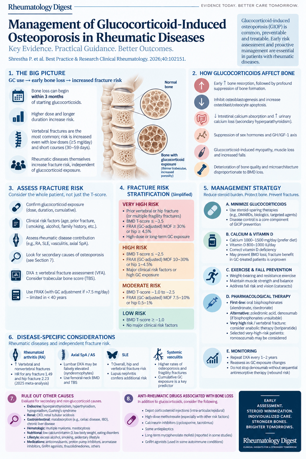
TL;DR: Glucocorticoids degrade bone quality faster than they lower bone density, so fracture risk must be assessed within three months of starting steroids — and a reassuring DXA does not rule it out.
Glucocorticoid-induced osteoporosis is the most predictable complication in rheumatology and still one of the least reliably acted on. The reason is partly structural: the tool most of us reach for to decide whether to treat is the one that performs worst in exactly this setting. This review sets out how quickly bone is lost, why the T-score misleads, how much the underlying disease adds on top of the drug, and what the evidence actually supports doing about it.
The damage starts early and tracks both dose and duration
Long-term glucocorticoid use is the leading cause of secondary osteoporosis. Despite the arrival and wide availability of steroid-sparing therapies, oral glucocorticoid use for more than three months in rheumatic disease has continually increased over the last twenty years.
Bone mineral density loss begins within three months of starting steroids and mainly affects trabecular-rich sites such as the lumbar spine, though cortical bone is also affected. Loss is dose- and duration-dependent, with higher doses producing greater losses, and exposure beyond ninety days associated with increased fracture risk and further BMD loss. Changes in bone microarchitecture are likewise dose-dependent and can appear within as little as three months of administration. The thoracic and lumbar vertebral column is the most common site of osteoporotic fracture in long-term users; sites with predominantly cortical bone, such as the distal forearm, are less commonly involved.
On the fracture side, risk rises in a dose- and duration-dependent manner, and is elevated even at low daily doses and within the first few months. Prednisone equivalents of ≥7.5 mg/day confer substantially higher fracture risk, with up to a fivefold increase for vertebral fractures. Duration is the single most important determinant, since BMD falls most rapidly during the first three to six months: fracture risk increases by approximately 75% in the first three months and continues to rise with ongoing treatment.
The thresholds are lower than most of us carry in our heads. Even short-term daily doses of ≥5 mg prednisone equivalents for thirty to fifty-nine days elevate vertebral and hip fracture risk, and doses of ≥1 and <2.5 mg daily for ninety days significantly increase vertebral and hip fracture risk. There is no comfortable dose.
Routes other than the daily tablet
Data on intravenous pulse steroids are limited. In a cohort of RA patients receiving IV pulse therapy, BMD at the lumbar spine, femoral neck and total body was not significantly reduced compared with baseline, whereas BMD was significantly decreased at all sites examined in patients treated with oral methylprednisolone. Against that, in patients with Graves’ orbitopathy treated with IV pulse therapy, even a single dose decreased bone formation and resorption markers.
Epidural administration has historically produced inconsistent reports, but more recent data suggest the frequency and total number of epidural steroid injections negatively affect BMD. A study of people receiving epidural injections for radicular back pain observed a 39% higher fracture rate among those who received them compared with those who did not, and rates of spine fractures — an area particularly vulnerable to steroid effects — were 54% higher. For intra-articular administration, suppression of bone formation has been reported, but no studies report changes in BMD or fracture risk.
What happens after the steroids stop
Recovery of BMD after glucocorticoids is gradual and often incomplete, and cumulative exposure predicts fracture risk after discontinuation. One study reported that fracture risk returned to baseline six months after discontinuation in those with under 1 g cumulative dose, whereas those with ≥1 g did not return to baseline until fifteen months later. There is also a genetic contribution to steroid-related bone loss and fracture risk.
The practical implication the authors draw is one that is easy to miss: it may be appropriate to consider continuing fracture prevention therapy beyond glucocorticoid discontinuation for a finite period, on the order of six to twelve months — even in patients who appear to be at low fracture risk at the point of stopping.
Why glucocorticoid bone is different
The glucocorticoid receptor sits in the cytosol of most cells complexed with heat shock proteins. Cortisol diffuses through the cell membrane and binds it, releasing those proteins; the activated receptor is transported into the nucleus, where it either attaches directly to DNA and stimulates gene expression, or complexes with other transcription factors and prevents them binding — the latter being the mechanism that suppresses inflammatory genes normally upregulated by NF-κB or AP-1.
The dominant skeletal effect is impairment of bone formation: glucocorticoids inhibit the proliferation and differentiation of osteoblasts and enhance apoptosis of osteoblasts and osteocytes. Several extra-osseous mechanisms compound this. Steroids decrease calcium-binding protein in the intestine, lowering calcium absorption and bone mineralisation, while increased bone resorption may cause excessive urinary calcium excretion and trigger secondary hyperparathyroidism. At high doses they reduce not only ACTH but LH and FSH, lowering estradiol, estrone, DHEAS and testosterone. Growth hormone, IGF-1 and IGF-binding protein levels are reduced by prolonged use, and steroids increase IGFBP transcription, which decreases IGF2, a local regulator of osteoblast function. High doses also reduce protein synthesis and cause loss of muscle mass. Polymorphisms in the glucocorticoid receptor have been linked to varying responses and to fracture risk, which may explain some of the individual variation.
The histology is the part worth remembering, because it explains the therapeutic logic. Remodelling is initially activated, producing high bone resorption; over time resorption parameters fall and bone becomes quiescent. Biopsies from patients on steroids for more than twelve months show increased resorption, decreased formation and decreased trabecular volume. Crucially, GIOP differs structurally from postmenopausal osteoporosis: in GIOP the trabeculae are reduced in number and surface area and are thin, but still connected, whereas in postmenopausal osteoporosis trabeculae are perforated by resorption with loss of continuity. The preservation of thinned but intact trabeculae in GIOP may provide the scaffold on which new bone can be formed — which is the argument for anabolic therapy.
What the underlying disease adds
Fracture risk in these patients is not attributable to the drug alone, and the contributions differ by disease.
Rheumatoid arthritis carries a 1.5- to 3-fold increased risk of vertebral and nonvertebral fractures across all fracture sites, with a comparable elevation in both sexes. A 2025 meta-analysis of twenty-nine prospective cohorts reported a hazard ratio of 1.49 (95% CI 1.35–1.65) for any clinical fracture and 2.23 (95% CI 1.85–2.69) for hip fracture — independent of sex, BMD and glucocorticoid exposure.
Ankylosing spondylitis presents a measurement problem rather than just a risk problem. Patients are at increased risk of vertebral fractures, but BMD of the lumbar spine is difficult to assess accurately by DXA: BMD is decreased at the hip but not at the lumbar spine later in the disease course, a paradox caused by bridging syndesmophytes and ligamentous ossification that artificially inflate the DXA reading. Trabecular bone score — a grey-level textural measurement derived from standard lumbar spine DXA images (L1–L4), where higher values correlate with better microstructure — helps here. Low TBS (<1.23) is associated with a 5.3-fold increased risk of vertebral fracture (OR 5.3, 95% CI 2.0–14.1), and the presence of syndesmophytes independently increases the odds of vertebral fracture (OR 5.5, 95% CI 2.2–13.5). Femoral neck BMD and TBS are therefore more accurate predictors than lumbar spine BMD in this group.
SLE carries elevated fracture risk across several skeletal sites (RR 1.97, 95% CI 1.20–3.25), particularly hip (RR 1.99, 95% CI 1.55–2.57) and vertebral fractures (RR 2.97, 95% CI 1.71–5.16). Risk of fragility fracture and osteoporosis is higher in those with lupus nephritis.
Psoriatic arthritis and psoriasis show inconsistent findings for osteoporosis and osteopenia, but patients nonetheless have a higher fracture risk than controls.
PMR and giant cell arteritis are difficult to disentangle from steroid exposure, since glucocorticoids are first-line for both. In a Swiss population-based study, osteoporosis was almost three times more common in GCA than in the general population.
Systemic vasculitis is similar, with high-dose IV steroids often required initially. A meta-analysis of forty studies reported a pooled osteoporosis prevalence of 14.6% (95% CI 12.2–18.9) and fragility fracture prevalence of 17.1% (95% CI 11.4–24.8). Against healthy controls, the odds ratio was 2.92 (95% CI 1.72–4.98) for osteoporosis and 2.39 (95% CI 1.34–4.26) for fragility fracture, with cumulative glucocorticoid exposure a significant predictor.
Secondary causes are common enough to look for routinely
Screening for secondary causes is needed in all patients at risk of fracture, including those on steroids — and the yield is higher than expected. Up to 30% of postmenopausal women, more than 50% of premenopausal women, and between 50% and 80% of men have a secondary cause of osteoporosis. Candidate causes include endocrine, neuromuscular and gastrointestinal disease, chronic inflammatory conditions, chronic kidney disease, nutritional impairment, organ transplantation, HIV, malignancy and genetic conditions. Response to anti-osteoporosis treatment may be suboptimal if the underlying condition is unrecognised and untreated.
The suggested workup is: serum calcium (corrected for albumin) or ionised calcium, phosphate, creatinine with estimated GFR, alkaline phosphatase, liver function tests, 25-hydroxyvitamin D, complete blood count, serum protein electrophoresis in older individuals, and testosterone in men. A 24-hour urine calcium collection should be obtained to assess the adequacy of calcium intake and absorption, with urinary creatinine in the same collection to confirm completeness — note that a spot urine calcium/creatinine does not accurately detect hypercalciuria. Urinary sodium is useful where hypercalciuria is suspected, since high sodium intake increases urinary calcium losses.
Two contributors deserve specific attention. Vitamin D deficiency impairs calcium absorption and bone mineralisation. And chronic kidney disease disrupts calcium and phosphate homeostasis: advanced CKD carries up to an 8-fold higher fracture risk than the general population, with fracture incidence per 1000 person-years rising progressively at 15.0, 20.5, 24.2, 31.2 and 46.3 across CKD stages 1–2, 3a, 3b and 4.
The rest of the medication list
Several drugs on a rheumatology chart influence bone, in both directions.
Methotrexate is reassuring at rheumatological doses. Standard low-dose methotrexate (<25 mg/week) has not been associated with BMD loss in large cohort and cross-sectional studies in RA and other inflammatory diseases, and therapy with methotrexate, sulfasalazine or hydroxychloroquine alone or in combination is not associated with incident fractures in postmenopausal women with RA.
Opioids have at most a short-term, carefully circumscribed role, since long-term use produces no improvement in function or pain control in rheumatic disease. Fracture risk is increased, highest in the early stages of use and with higher doses, with adjusted odds ratios ranging from 1.5 to 2.7.
Antidepressants have been associated with significant BMD reduction at multiple sites and a 2.5-fold increased risk of hip fracture in users compared with non-users. There are, however, no data suggesting duloxetine — the SNRI most used in rheumatology for fibromyalgia — has any effect on skeletal health.
Gabapentinoids can mechanistically interfere with calcium channel function in bone and muscle. Clinical studies do not show significant differences in lumbar or femoral neck BMD or bone turnover markers between pregabalin users and controls, though there may be a trend toward lower lumbar spine BMD in men and in those treated for less than twenty-four months.
Calcineurin inhibitors matter because calcineurin is a phosphatase critical for osteoblast differentiation and bone formation. Tacrolimus is linked to increased bone resorption and lower BMD, with higher blood concentrations correlating with greater bone loss; the risk is dose-dependent and more pronounced with longer use. For voclosporin, no studies have specifically evaluated bone metabolism or fracture risk in steroid-treated patients — AURA-LV and AURORA 1 used rapidly tapered steroid regimens but did not report bone-specific outcomes.
Reproductive health medications are easy to overlook. Leuprolide induces hypogonadism, with BMD loss of 4–6% at the lumbar spine after six months, partially recovering after discontinuation. Depot medroxyprogesterone acetate suppresses estrogen production, producing BMD loss of 0.5–3.5% at hip and spine after one year and up to 5–7% after two years; it carries an FDA boxed warning for significant, potentially irreversible bone loss with prolonged use, especially in adolescents and young women, and recovery after discontinuation is incomplete, particularly with longer use.
Why the T-score misleads, and what TBS adds
This is the single most clinically important point in the review.
DXA with vertebral fracture assessment where possible should be initiated as soon as possible after starting prednisone at ≥2.5 mg/day or equivalent, and BMD with VFA or spinal x-rays is also advised in patients under forty.
But: BMD measurements by DXA frequently underestimate fracture risk in GIOP, because glucocorticoids disproportionately impair bone quality and microarchitecture — especially at trabecular-rich sites — without always causing marked reductions in density. Patients on long-term steroids can sustain fragility fractures at BMD values above the conventional osteoporosis threshold of T-score > −2.5. A normal or near-normal DXA in a patient on steroids is therefore not the reassurance it appears to be, and height loss remains an important clinical indicator of possible vertebral fractures.
Trabecular bone score helps close the gap. TBS is more sensitive than areal BMD for fracture detection in GIOP, and adding it to BMD substantially improves fracture risk stratification. It is most useful as an adjunct to DXA in precisely the patients where DXA is most misleading: those whose BMD is normal or only mildly reduced but who carry significant clinical risk factors.
FRAX, and the blind spot it has for steroids
For adults aged forty and over, FRAX is the most commonly used tool and incorporates glucocorticoid use as a risk factor. The assumed exposure built into the calculator is a moderate dose of 2.5–7.5 mg prednisolone daily or equivalent, and it can be adjusted upward: for doses >7.5 mg/day, multiply the 10-year major osteoporotic fracture risk by 1.15 and the hip fracture risk by 1.2.
The limitation is structural — FRAX does not account for cumulative glucocorticoid dose and may therefore underestimate risk in some patients. The FRAX plus tool allows imputation of steroid doses but requires a fee. QFracture includes glucocorticoids as an individual risk but does not adjust for dose. And in adults under forty, FRAX cannot be used at all: risk assessment there rests mainly on BMD and prior fracture history, with vertebral fracture assessment where appropriate.
The first intervention is using less steroid
Before any bone-directed drug, the ACR recommends prioritising strategies that reduce glucocorticoid burden — tapering and switching to steroid-sparing regimens as disease control allows. The European Calcified Tissue Society makes the same argument from the other direction: fracture risk is highest early in the course of therapy, so optimal management of the underlying disease, often via steroid-sparing agents, reduces the need for prolonged or high-dose exposure. The review then goes disease by disease.
SLE. The 2025 ACR guideline emphasises hydroxychloroquine, limitation of steroid exposure, and early introduction of immunosuppressive therapy to achieve remission or low disease activity. For stable patients on >5 mg/day prednisone, tapering to ≤5 mg/day and ideally discontinuing within six months is recommended. In lupus nephritis, early combination therapy with mycophenolate mofetil, cyclophosphamide or calcineurin inhibitors allows steroid tapering. Notably, short-term pulse IV methylprednisolone for severe organ involvement permits more rapid oral reduction, and the cumulative dose from pulse therapy is not considered equivalent to chronic high-dose oral exposure in terms of osteoporosis risk. In proliferative (class III/IV) disease, induction with pulse IV steroids plus mycophenolate or cyclophosphamide is followed by maintenance, targeting oral prednisone ≤7.5 mg/day by three to six months.
Giant cell arteritis. IV methylprednisolone (125–500 mg/day for three days) plus methotrexate with lower-dose oral prednisone (≤30 mg/day) achieves similar remission and relapse rates to standard high-dose oral prednisone (40–60 mg/day), but with substantially lower cumulative exposure, a much faster taper to ≤5 mg/day (mean 13.8 versus 56.5 weeks) and significantly lower risk of steroid-related adverse effects. In the GiACTA trial, sustained remission at week 52 was achieved in 56% on weekly tocilizumab plus a 26-week prednisone taper, versus only 14–18% on placebo with the standard taper, and tocilizumab reduced median cumulative prednisone over 52 weeks by nearly half. In a randomised trial, upadacitinib 15 mg once daily significantly reduced cumulative glucocorticoid exposure over one year.
Polymyalgia rheumatica. Sarilumab is FDA-approved for patients with an inadequate response to steroids or who cannot tolerate a taper; sarilumab plus a 14-week prednisone taper produced significantly higher sustained remission rates and lower cumulative doses than placebo.
ANCA-associated vasculitis. Reduced-dose regimens and early steroid-sparing agents — rituximab, cyclophosphamide, avacopan — should be considered, with PEXIVAS and ADVOCATE having established lower-dose approaches as standard of care. Avacopan was noninferior to prednisone for remission at 26 weeks and superior for sustained remission at 52 weeks, with significantly lower glucocorticoid toxicity scores and fewer serious adverse events; subgroup analyses show the greatest benefit in patients at high risk of steroid toxicity, including older age, diabetes, osteoporosis and infection risk. PEXIVAS reported that a reduced-dose regimen — roughly 50% lower cumulative exposure over six months — was noninferior to standard dosing, with significantly lower risk of steroid-related adverse events and without adversely affecting remission or survival.
Calcium, vitamin D, protein and exercise
The evidence here is weaker than the confidence with which it is usually prescribed, and the review is refreshingly direct about it. Most studies of calcium and vitamin D supplementation were conducted in community-dwelling people, not specifically in those treated with glucocorticoids.
A large meta-analysis concluded that increasing calcium intake, by diet or supplement, produced a benefit of about 1% on BMD at the end of the first year, which did not increase further with continued supplementation — independent of whether vitamin D was given, and unrelated to baseline calcium intake or dose. Most studies show no relationship between calcium intake and fracture risk; one meta-analysis found calcium monotherapy tended to increase hip fracture risk (RR 1.53; 95% CI 0.97–2.42) and had no effect when combined with vitamin D. Adding calcium supplements to anti-resorptive drugs has not been shown to substantially affect fracture risk. Supplementation is best achieved through diet, which does not carry the constipation, renal stone and possible cardiovascular risks of supplements.
Vitamin D supplementation may prevent BMD loss in those who are deficient, and correction of deficiency (<25 nmol/L) is necessary before starting anti-resorptive drugs to avoid hypocalcaemia — a practical sequencing point. Most recommendations target a minimum 25-hydroxyvitamin D of 30 ng/ml. In the one meta-analysis that did include steroid-treated patients, there was significant prevention of BMD loss at lumbar spine and forearm with calcium and vitamin D — but there are no data showing that calcium and/or vitamin D prevent fractures in people taking glucocorticoids. The ACR nonetheless recommends all patients on steroids take at least 1500 mg calcium and 800 IU vitamin D daily, preferably through diet where possible.
On protein, older adults with chronic illness are advised to consume ≥1.2–1.5 g/kg/day; a systematic review found intake above the 0.8 g/kg/day RDA associated with reduced hip fracture risk and positive trends in total hip and femoral neck BMD — though this was not studied in GIOP.
Exercise data are limited. A systematic review and meta-analysis including three trials over six months found a significant positive effect of exercise on lumbar spine BMD but not hip. Falls increase rapidly after steroid initiation and return to baseline fairly rapidly after discontinuation, and steroids also increase cataract risk, which may impair visual acuity and further increase falls. No study has directly examined whether fall prevention reduces fragility fractures in patients receiving glucocorticoids. Low-intensity vibration therapy has recently been FDA-approved and reports improvements in lumbar and hip BMD and muscle strength, but protocols are not standardised and there are no data in GIOP.
What the drug trials actually show
Bisphosphonates are the most studied class, with alendronate most common, followed by risedronate, zoledronic acid, teriparatide, denosumab and romosozumab. The trials differ substantially in design — demographics and comorbidities, whether calcium and vitamin D were given and at what dose, whether rheumatic disease patients were included and which ones, disease severity, average prednisone dose and duration, trial length and outcomes assessed. That heterogeneity is the reason the comparative picture stays blurry.
Placebo-controlled trials, which would not be considered ethical today, reported improvements in BMD and/or fracture reduction with active drug. Alendronate produced significant BMD increases at multiple sites including lumbar spine and femoral neck, with Saag (1998) reporting a reduction in new vertebral fractures and Gonnelli reporting improved BMD at lumbar spine, femoral neck and trochanter with reduced incident vertebral fractures. Yilmaz (2001) showed lumbar spine BMD increased significantly with alendronate versus no treatment. For risedronate, Reid (2001) showed significantly improved BMD at lumbar spine, femoral neck and trochanter with reduced incident vertebral fractures, and Eastell (2000) reported a significant difference versus placebo at the lumbar spine (1.4% versus −1.6%) and trochanter (−0.4% versus −4%). Alendronate consistently suppressed bone turnover markers across trials.
Active comparator trials show differences between agents. Romosozumab was associated with higher lumbar spine BMD gains than denosumab. Denosumab demonstrated superior lumbar spine BMD improvement compared with alendronate, and Mok (2015) showed that switching to denosumab provided higher lumbar spine BMD gains and stronger bone turnover suppression than continuing oral bisphosphonates. Among anabolics, teriparatide consistently outperformed alendronate at lumbar spine and hip and reduced vertebral fractures — Saag reported a lumbar spine increase of +10.3% with teriparatide versus +5.5% with alendronate. Among bisphosphonates, zoledronic acid had a stronger antiresorptive effect and greater lumbar spine and hip gains than risedronate, while in another study alendronate was associated with fewer fractures than risedronate.
The honest summary is the important one. FDA-approved osteoporosis drugs have shown efficacy in preventing and treating GIOP, but few report vertebral fracture data and none have reported hip or nonvertebral fracture reduction. All demonstrate benefit over placebo for BMD change and, in some cases, reduce vertebral fracture risk. Because of the variability between study populations and designs, it is not possible to confidently rank superiority across drug classes. What the literature does support is that anabolic agents produce larger BMD gains in GIOP, and that in high-risk populations anabolic agents should be prioritised over antiresorptives — consistent with the histological picture of thinned but preserved trabeculae.
On the individual agents: oral bisphosphonates (alendronate, risedronate) are antiresorptive and generally first-line for moderate-to-high risk, with GI irritation the main caution and severe CKD a reason to avoid. Zoledronic acid is the IV option when oral agents are not tolerated, with acute phase reaction, renal toxicity and hypocalcaemia to watch. Teriparatide, a PTH analogue, is anabolic and preferred in very high risk, rapid bone loss or vertebral fracture, with hypercalcaemia the main precaution. Denosumab, a RANKL inhibitor, is an alternative where bisphosphonates are unsuitable but carries the well-known risk of rebound fractures if stopped without transition therapy, plus hypocalcaemia. Romosozumab, a sclerostin inhibitor with both anabolic and antiresorptive action, is reserved for very high-risk patients where other agents are inadequate or contraindicated; it carries an FDA black box warning for increased cardiovascular risk and requires transition therapy after twelve months. Atypical femoral fractures and medication-related osteonecrosis of the jaw are recognised late sequelae of this drug class generally.
Where the guidelines converge
Guidance from the ACR, ECTS, Japanese, UK, Colombian and Latin American societies agrees more than it differs.
Assess early. All major guidelines treat early, accurate fracture risk assessment as the cornerstone. Patients receiving ≥3 months of systemic glucocorticoids should undergo fracture risk assessment at the initiation of therapy, with reassessment during treatment. The ACR recommends FRAX adjusted for steroid dose, validated in adults over forty, combined with BMD. Japanese guidelines integrate clinical risk factors — age, sex, prior fracture, fall risk, serum calcium, vitamin D and steroid dose — with BMD-based decision-making, and ECTS supports the same combined assessment. Colombian and European/Belgian frameworks emphasise individualised assessment accounting for comorbidities and local resource availability.
Treat at moderate risk and above. Pharmacologic treatment is strongly indicated for moderate or high fracture risk, including prior fragility fracture, T-score ≤ −2.5, or elevated FRAX adjusted for steroid dose. High-risk patients — a FRAX-derived major osteoporotic fracture risk ≥10% or prior fragility fracture — require pharmacologic therapy, while low-risk individuals may benefit primarily from non-pharmacologic strategies. The ACR separately identifies cumulative exposure >5 g/year of prednisone equivalents, particularly combined with very high daily doses of ≥30 mg/day, as a threshold warranting pharmacologic intervention.
Monitor and plan the exit. Regular BMD monitoring every one to two years is recommended by the ACR, ECTS and UK guidelines. Treatment duration should align with the dose and duration of steroid exposure, with reassessment when steroids are tapered or stopped. Japanese and European guidelines highlight the need for vigilance about rebound bone loss after stopping denosumab. Across guidelines, non-pharmacological recommendations settle on calcium 1000–1200 mg/day and vitamin D 800–1000 IU/day, with weight-bearing exercise, fall prevention and lifestyle modification.
What remains unsettled
The research agenda is specific about the gaps. Randomised trials are needed comparing bisphosphonates, denosumab and anabolic agents — including newer anabolics such as abaloparatide and romosozumab — for fracture prevention in GIOP in people with rheumatic disease. And observational studies using a targeted trial emulation approach are needed to understand the comparative efficacy and safety of sequential therapies, which is how these drugs are actually used.
The authors’ practice points are worth restating plainly. All adults receiving three months or more of systemic glucocorticoids should undergo early fracture risk assessment. Patients on long-term steroids can sustain fragility fractures with BMD above the conventional threshold, because BMD underestimates risk in this setting. Early consideration of steroid-sparing agents for the underlying rheumatic disease is important. And among current FDA-approved therapies, teriparatide has the strongest data for BMD increase and vertebral fracture reduction.
Read together, the message is that the sequence most of us follow — start steroids, order a DXA at some point, act if the T-score is bad — inverts the actual evidence. The assessment belongs in the first three months, the steroid-sparing conversation belongs at initiation, and the T-score is the least trustworthy input in the whole decision.
Funding noted in the source: supported by grant R01AG079118 from the National Institute on Aging. The authors declare no competing interests.