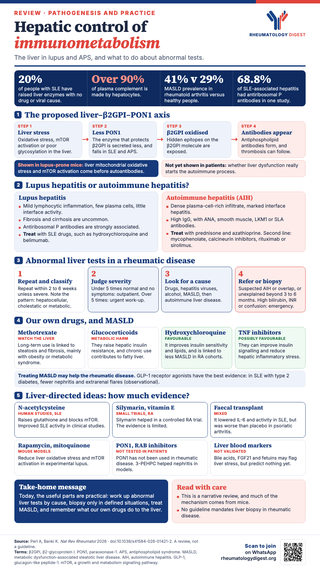
TL;DR: This review proposes that the liver sits upstream of autoimmunity in lupus and antiphospholipid syndrome, because hepatocytes make the antigen (β2-glycoprotein I) and the enzyme that protects it from oxidation (PON1), and in lupus-prone mice hepatic oxidative stress and mTOR activation come first, but most of the mechanism is preclinical, while the clinically usable parts are a structured work-up of abnormal liver tests, selective biopsy, awareness of MASLD, and the liver effects of our own drugs.
Autoimmune rheumatic diseases (ARDs) are usually studied from the side of the immune system. This review, by Andras Perl and Katalin Banki (SUNY Upstate Medical University), asks what the liver contributes. It is a narrative review rather than a systematic one, and a good deal of it rests on mouse models, genetic deficiencies and small studies, so a reader should treat the central idea as a well-argued hypothesis, not as an established sequence of events. The paper’s key points are that the liver integrates metabolic and immune signalling, that hepatic mitochondrial oxidative stress and mTOR activation precede autoimmunity in lupus-prone mice, that hepatocyte-derived β2-glycoprotein I (β2GPI) is oxidatively modified into an immunogenic form, that liver-derived paraoxonase-1 (PON1) protects it, and that intrahepatic immunometabolic pathways are potential treatment targets.
What the liver does for the immune system
The liver is the main metabolic organ and a major immune organ. Its cell types each have an immune role:
- Hepatocytes make complement, coagulation factors, acute-phase proteins, lipoproteins, β2GPI (apolipoprotein H) and PON1, and metabolise nutrients, drugs and toxins.
- Kupffer cells are the liver’s resident macrophages. They account for 80–90% of tissue macrophages in the body, filter bacteria and debris from portal blood, clear immune complexes (which build up in SLE and vasculitis) and make IL-10, which supports regulatory T-cell development.
- Liver sinusoidal endothelial cells (LSECs) expand TGFβ-dependent regulatory T cells and express inhibitory molecules such as PD-L1, which supports tolerance.
- Stellate cells store vitamin A (80–90% of the body’s stores), and when activated become collagen-producing cells that drive fibrosis and make IL-6 and TGFβ.
About 70% of the liver’s blood supply comes through the portal vein, so it is constantly exposed to gut-derived microbial products, metabolites and dietary antigens. The authors describe it as a tolerogenic organ that decides whether these signals lead to tolerance or to systemic inflammation.
Development. The liver is the main site of haematopoiesis during fetal life, and hepatic stromal cells and hepatocytes supply growth factors and cytokines (stem cell factor, IL-7, thrombopoietin, FLT3 ligand, IL-6, IL-4 and IL-15 for natural killer cells). The authors suggest that disrupted hepatic metabolism in fetal life, through genetic defects that alter oxidative stress or glycosylation, can have long-lasting immunological effects.
Liver-made proteins that matter in rheumatology
- Complement. Hepatocytes make over 90% of plasma complement, including C3, C4, C5, C6, C8, C9, factors B, H and I and C1 inhibitor. C1q, factor D, properdin and C7 come mostly from other cells. Inherited deficiency of the liver-made classical components C2 and C4 markedly raises susceptibility to autoimmune disease, especially SLE.
- CRP and other acute-phase proteins. CRP is made by the liver in response to IL-6 and other cytokines. The circulating pentamer is generally inert or weakly anti-inflammatory, while the monomeric form can deposit in vessel walls, recruit macrophages, activate platelets and promote foam-cell formation. The authors raise the possibility that liver-derived CRP contributes to the roughly fivefold higher cardiovascular mortality in ARDs. Genetic variants that lower CRP expression predispose to SLE and RA, and CRP also helps clear apoptotic cells. Serum amyloid P limits fibrosis, serum amyloid A activates neutrophils, macrophages and dendritic cells, and ferritin promotes release of IL-1β, IL-6 and TNF.
- Iron and hepcidin. Hepcidin, induced by IL-6, is the master regulator of iron. It binds ferroportin, causing its degradation, traps iron in macrophages and lowers dietary absorption, which gives an iron-restricted anaemia that resists oral iron. Plasma hepcidin is raised in SLE, and in mice hepcidin reduced renal inflammation in lupus nephritis. Hereditary haemochromatosis, from HFE mutations that limit hepcidin, causes iron overload with chondrocalcinosis and inflammatory arthritis.
- Alpha-1 antitrypsin (AAT). About 90% comes from hepatocytes. Its deficiency causes uncontrolled neutrophil elastase activity with injury to lung and kidney, promotes anti-citrullinated protein antibody production and disease severity in RA, and is linked to granulomatosis with polyangiitis (AAT inhibits proteinase 3, the main ANCA target). Direct AAT administration might help in SLE, according to the authors.
- Mannose-binding lectin (MBL). Hepatocytes are the main source. Deficiency predisposes to SLE and juvenile RA, although levels are often raised in established SLE, possibly as compensation.
- HMG-CoA reductase. Mostly expressed in the liver, where statins act. Statins also induce abnormal expression of the enzyme in muscle and can trigger anti-HMG-CoA autoantibodies and autoimmune myositis.
- Coagulation and thrombopoietin. Hepatocytes make the clotting factors and the anticoagulants antithrombin, protein C, protein S and protein Z. Autoantibodies against these anticoagulants are associated with thrombosis in SLE. Thrombopoietin is also made in the liver, and autoantibodies to it and its receptor occur in about one-third of people with SLE, especially with thrombocytopenia. The authors note that vitamin K antagonism with warfarin remains the optimal preventive treatment for APS, and that variants in the carboxylase enzyme (GGCX) can affect how well it works.
Gut, lung and skin signals pass through the liver
Hepatocytes turn cholesterol into primary bile acids, gut bacteria convert them into secondary bile acids, and these shape immunity:
- Taurochenodeoxycholic acid acting on the TGR5 receptor pushes monocytes towards IL-12-poor, tolerogenic dendritic cells.
- Deoxycholic acid dampens inflammatory dendritic cell responses in experimental autoimmune models through the TGR5–cAMP–PKA pathway.
- Lithocholic acid derivatives (3-oxoLCA, isoalloLCA) suppress TH17 cells and promote regulatory T cells.
For the lung, isolithocholic acid made from liver-derived bile acids suppresses cytokine production by pulmonary macrophages in vivo, although the authors state that direct evidence of the liver changing the lung microbiome is lacking. For skin, bile acids act on keratinocytes through farnesoid X receptor signalling and influence antimicrobial peptides, with proposed relevance to rosacea and psoriasis. The skin does not drain into the liver through a portal circulation, but skin-derived mediators and metabolites enter the systemic circulation and are processed by the liver.
How the liver may turn self into autoantigen
Hepatocytes carry out extensive post-translational modification of proteins and lipids, and the review argues this can convert normal molecules into autoantigens.
- Oxidation. β2GPI normally circulates in a closed conformation that hides its immunogenic epitopes, and oxidation exposes cryptic epitopes recognised by antiphospholipid antibodies. Oxidative DNA damage in hepatocytes produces modified bases (8-hydroxy-2′-deoxyguanosine and 5-hydroxymethylcytosine, the latter made by TET2) that are linked to ANA production. In mice, a high-fat diet and glutathione deficiency raise TET2 in hepatocytes.
- Glycosylation. PON1 needs glycosylation for secretion. In transaldolase (TAL) deficiency, carbon becomes trapped in the pentose phosphate pathway, UDP-GlcNAc is depleted, PON1 secretion falls and anti-β2GPI and anticardiolipin antibodies appear. The metabolic picture (glutathione depletion, kynurenine accumulation) resembles SLE. Glycosylation also matters for complement factors and for AAT’s ability to break down immune complexes.
- Lipidation. β2GPI binds negatively charged phospholipids such as cardiolipin and phosphatidylserine. Cardiolipin becomes antigenic when apoptosis and oxidative stress rise in the livers of lupus-prone mice, and oxidised lipids can change phospholipid composition in ways that favour β2GPI binding.
- Citrullination and other changes. Stellate cells express PAD2, people with autoimmune hepatitis can develop anti-CCP antibodies, and loss of TNFAIP3 (A20) leads to PAD4 overexpression in SLE and also predisposes to MASH.
- NAD supply. About 95% of circulating nicotinamide is released by the liver, so liver-derived NAD probably affects immune cell metabolism, from mitochondrial function to sirtuin-dependent epigenetic marks.
The liver–β2GPI–PON1 axis
This is the core proposal. Both β2GPI and PON1 are expressed only by hepatocytes and secreted into the blood. PON1 protects lipoproteins and β2GPI from oxidative modification, and considerable loss of PON1 activity is seen in APS and SLE. The sequence the authors propose: hepatic metabolic defects (impaired glycosylation or oxidative stress) lower PON1 secretion, β2GPI is oxidised and becomes immunogenic, and antiphospholipid antibodies appear. Antiphospholipid antibodies often appear before clinical disease, and APS and SLE can show liver dysfunction before overt autoimmunity.
Evidence from mice. In lupus-prone MRL-lpr mice, mitochondrial oxidative stress and mTOR activation in the liver precede autoantibody production. Overexpression of RAB4A drives the increased mitochondrial mass and ROS, and antioxidants such as N-acetylcysteine reduce these abnormalities and delay disease. Evidence in people is more limited: about 20% of people with SLE have liver injury unrelated to immunosuppressive treatment or viral infection, autoimmune hepatitis can precede SLE, and PON1 activity is reduced in SLE and APS. The review concludes that hepatic autoimmunity might be an early pathogenic event in SLE, not merely a consequence, but this has not been shown in patients.
Other liver-derived protection. The liver is the main source of glutathione, albumin (which supplies about 80% of the blood thiol pool through its Cys34 residue), bilirubin, selenoprotein P (which protects against ferroptosis, including in lupus nephritis kidneys) and ceruloplasmin (which carries over 95% of plasma copper). PON1 and PON3 protect LDL and HDL from oxidation, and low PON1 is associated with atherosclerosis in SLE and RA. PON1 also hydrolyses homocysteine thiolactone, and its deficiency leads to N-homocysteinylation of fibrinogen and other coagulation proteins, which the authors link to thrombosis.
Kynurenine, drugs and toxicants
Kynurenine is the most accumulated metabolite in SLE and Sjögren disease and drives inflammatory T-cell development in SLE. Five possible sources are proposed: TAL deficiency; liver-resident Enterococcus gallinarum converting tryptophan; RAB4A overexpression promoting uptake; dietary tryptophan (a low-tryptophan diet limited kynurenine build-up, TH17 development and nephritis in lupus-prone mice); and raised TDO or IDO enzyme activity. TDO is expressed mainly in the liver.
Drug metabolism. Procainamide and hydralazine can cause drug-induced lupus through DNA hypomethylation. Procainamide is cleared by hepatic N-acetylation, and slow acetylators have about four-fold higher risk of ANA and SLE. The acetylated metabolite has little propensity to cause lupus. Toxicants metabolised by the liver, including heavy metals, pesticides and air pollutants, have been implicated in SLE and systemic sclerosis, because reactive intermediates can cause oxidative stress.
Liver involvement in specific rheumatic diseases
SLE. Around 20% of patients have raised liver enzymes independent of drugs or viral infection, and lupus hepatitis can be the first manifestation. Lupus hepatitis and autoimmune hepatitis (AIH) are distinguished as follows:
- Lupus hepatitis: usually a non-specific reactive hepatitis with mild portal and lobular lymphocytic inflammation, little or no interface activity and few plasma cells. Substantial fibrosis and cirrhosis are uncommon, and histology tends to track overall SLE activity. Diffuse lobular inflammation and nodular regenerative hyperplasia (attributed to immune-complex-triggered obliterative venopathy) are typical, and antiribosomal P antibodies are strongly associated (present in 68.8% of SLE-associated hepatitis in one study and absent in isolated AIH).
- AIH: dense lymphoplasmacytic portal infiltrates, marked interface hepatitis, hepatocyte rosettes and varying fibrosis. Type I (lupoid) AIH affects young women with marked hypergammaglobulinaemia and ANA, type II mainly children and Mediterranean populations with anti-LKM antibodies and ANA negativity, and type III is ANA and smooth-muscle-antibody positive with anti-soluble liver antigen.
- Treatment differs. Lupus hepatitis is managed with SLE-directed drugs such as hydroxychloroquine and belimumab. AIH is treated first with prednisone and azathioprine, with mycophenolate, calcineurin inhibitors, rituximab and sirolimus as second-line.
APS. Liver disease is mainly from thrombosis of the hepatic vasculature, which is the most affected organ. Small intrahepatic vessel occlusion causes nodular regenerative hyperplasia and Budd–Chiari syndrome. People with antiphospholipid antibodies are predisposed to progressive liver disease including cirrhosis, and cirrhosis has been associated with APS.
Other diseases.
- Systemic sclerosis: liver fibrosis is surprisingly rare.
- Primary biliary cholangitis (PBC): destruction of small bile ducts, with anti-mitochondrial antibody (against PDC-E2) as the hallmark. It often coexists with Sjögren disease and RA.
- Primary sclerosing cholangitis: occurs in about 2% of people with inflammatory bowel disease, and gut–liver interactions may contribute to arthritis.
- RA: MASLD prevalence is higher than in healthy people (41.2% versus 28.5%, incidence rate ratio 1.35, 95% CI 1.14–1.60), even after adjusting for metabolic and lifestyle factors. TNF, IL-6 and IL-1β promote hepatic insulin resistance, lipid accumulation and stellate cell activation.
- Spondyloarthritis: liver involvement tends to come with inflammatory bowel disease or MASLD.
- Osteoarthritis: increasingly seen as metabolically influenced, with MASLD and metabolic syndrome linked to OA independent of body weight.
MASLD in rheumatic disease
MASLD affects up to 30% of the global population and is a bidirectional comorbidity in ARDs, driven by chronic inflammation and metabolic dysfunction. In support of the bidirectional idea, treating MASLD may improve a coexisting ARD.
- GLP-1 receptor agonists have the strongest evidence. They improve steatosis, lower liver inflammation and cause weight loss, and signalling through cAMP–PKA and AMPK inhibits mTORC1 and NF-κB. In people with both SLE and type 2 diabetes, GLP-1RA use was associated with better cardiac outcomes and a lower risk of lupus nephritis and extrarenal flares (observational data). The authors state that prospective studies in rheumatic disease are strongly warranted.
- Pioglitazone improves steatosis and MASH histology, especially in diabetes, and may reduce disease activity and vascular inflammation in RA and SLE.
- Sex. Evidence for sex-specific hepatic manifestations is limited. Liver abnormalities in SLE track disease activity, drugs and metabolic liver disease, while male sex alone seems to add susceptibility to steatosis and fibrosis when MASLD is present.
How our own drugs affect the liver
Conventional therapies change hepatic metabolism, independent of overt hepatotoxicity:
- Methotrexate: long-term exposure is associated with steatosis and fibrosis, with obesity and metabolic syndrome as major cofactors. The pattern looks like altered hepatic lipid handling rather than direct hepatocyte injury alone, and electron microscopy shows collagen deposition in the space of Disse before clinically meaningful fibrosis.
- Hydroxychloroquine: improves insulin sensitivity, lipid profile and fasting glucose, is linked to lower diabetes risk, and to lower MASLD prevalence in RA cohorts.
- TNF inhibition: can improve insulin signalling and reduce hepatic inflammatory stress, since TNF is a major mediator of hepatic insulin resistance.
- Azathioprine: can lower glutathione and impair hepatocyte mitochondrial function.
- Leflunomide: inhibits dihydroorotate dehydrogenase, a mitochondrial enzyme.
- Glucocorticoids: promote hepatic insulin resistance and gluconeogenesis, and chronic exposure contributes to metabolic syndrome and fatty liver.
Abnormal liver tests: approach and when to biopsy
The review gives a stepwise approach, adapted from hepatology guidance because no ACR–EULAR guideline mandates biopsy in ARDs with abnormal liver tests:
- Confirm and classify. Repeat testing within 2–6 weeks unless the abnormality is severe, then classify the pattern: hepatocellular (ALT and AST), cholestatic (ALP and GGT) or metabolic.
- Triage by severity. ALT and AST under five times the upper limit of normal in an asymptomatic patient go to outpatient evaluation. Over five times means an urgent work-up. Raised bilirubin, a high INR or encephalopathy is an emergency.
- Look for the cause. Drug-induced injury (methotrexate, azathioprine, leflunomide, NSAIDs, biologics), viral hepatitis (HBV, HCV, EBV, CMV), alcohol, MASLD (BMI, diabetes, hypertension, FibroScan or FIB-4), and autoimmune liver disease (ANA, anti-mitochondrial, LKM1, ribosomal P and soluble liver antigen antibodies, IgG). For a cholestatic pattern, add ultrasound, MRI or MRCP.
- Refer to hepatology, with or without elastography and biopsy, for persistent unexplained abnormalities, suspected AIH or overlap syndrome, or advanced fibrosis.
The consensus is that biopsy should be selective:
- Strongly recommended when AIH is suspected (raised ALT or AST with raised IgG and positive ANA, smooth muscle, LKM1 or SLA antibodies).
- Recommended to separate lupus hepatitis from AIH when the diagnosis is uncertain, and for suspected AIH–PBC or AIH–PSC overlap.
- Consider for persistent unexplained ALT or AST elevation beyond 3–6 months, unexplained cholestatic enzymes, atypical PBC (including AMA-negative), and for vasculitic, granulomatous or infiltrative liver disease when the result will change management.
- Usually not indicated for mild or transient abnormalities with an identifiable cause such as drugs, viral hepatitis or MASLD. Address reversible causes first.
- Persistent abnormalities on methotrexate: non-invasive fibrosis testing is preferred over biopsy.
Prospective studies of biopsy in ARDs are still needed.
Biomarkers of hepatic metabolic stress
Conventional ALT and AST have limited prognostic value. Candidate early markers of hepatic metabolic dysfunction in people at risk of autoimmunity include bile acid metabolites (LCA, deoxycholic acid, TCDCA, which accumulate before overt liver injury), FGF21 (mitochondrial stress), selenoprotein P (redox imbalance), fetuin-A (innate activation through TLR4), fetuin-B (steatosis), β-hydroxybutyrate (ketogenesis, which blocks the NLRP3 inflammasome) and branched-chain amino acids (which activate mTOR but are not liver-specific). None of these has been validated as a predictor of rheumatic disease.
Liver-directed treatments
Most of what follows is experimental. The review lists the targets: mitochondrial oxidative stress, mTOR activation, steatosis and abnormal handling of gut microbial products.
- N-acetylcysteine (NAC): replenishes glutathione and inhibits mTOR. Clinical studies suggest improved SLE activity through reduced T-cell activation, and meta-analyses are said to back this up.
- Rapamycin, mitoquinone (mitochondria-targeted antioxidant) and metformin: promising in experimental models, and metformin is reported to have therapeutic benefit in SLE.
- PON1: not yet used to treat ARDs, though it protects against organophosphate toxicity, experimental colitis and ischaemic stroke.
- RAB inhibitors: 3-PEHPC (blocks RAB4A and RAB5A geranylgeranylation) improves lupus nephritis in models. The RAB7 inhibitor CID-1067700 reduces autoantibodies but promotes MASLD by impairing lipophagy.
- Hydroxychloroquine: acts on lysosomal acidification and mTOR, and its main cellular target is PPT1. Inhibiting PPT1 blocks TLR9 binding of self-DNA in models, but PPT1 blockade can lower HDL and promote atherosclerosis. The authors add that hydroxychloroquine is metabolised in the liver to active metabolites that can accumulate in ocular tissue and may contribute to retinal toxicity.
- Ursodiol: the standard bile-acid treatment for PBC, which also blocks mTOR in hepatocytes and the development of pro-inflammatory CD4 T cells, with potential benefit in overlapping autoimmune disease such as SLE.
- Mycophenolate and azathioprine: both prodrugs activated in the liver (mycophenolate to mycophenolic acid by hepatic esterases; azathioprine to 6-mercaptopurine and thioguanine nucleotides). Their effect is on purine synthesis in lymphocytes.
- Natural compounds: curcumin, silymarin, vitamin E, glutathione and polyphenols. Silymarin showed benefit in a controlled RA trial, vitamin E had analgesic effects in a placebo-controlled RA trial, and meta-analyses report polyphenols improving DAS28, CRP and ESR in RA. The authors describe the clinical evidence as limited and call for controlled trials.
- Gut microbiome: E. gallinarum translocation to the liver triggers lupus-like autoimmunity in lupus-prone mice, and its DNA has been recovered from liver biopsies of people with SLE. Faecal microbiota transplantation from healthy donors in SLE promoted short-chain fatty acids, lowered IL-6 and reduced disease activity. Probiotics improved DAS28 and CRP in RA over 8 weeks, though a meta-analysis showed only moderate benefit in RA and none in spondyloarthritis. A proof-of-concept randomised trial of faecal transplantation in PsA found it safe but inferior to placebo at 26 weeks.
What it adds up to
The review’s conclusion is that the liver is a central regulator of systemic immunity and that hepatic metabolic dysfunction, especially oxidative stress and impaired secretion of protective proteins such as PON1, might precede systemic autoimmunity in SLE and APS. It calls for work on the molecular link between hepatic metabolism and immune regulation, hepatic biomarkers that predict autoimmunity in longitudinal cohorts, liver-specific genetic studies, and liver-targeted treatments that could prevent or ameliorate autoimmunity.
For the clinic today, the supported points are narrower: abnormal liver tests in an ARD deserve a systematic search for drug, viral, metabolic and autoimmune causes, biopsy should be reserved for defined situations, MASLD is a real and treatable comorbidity, and our own drugs have measurable effects on the liver. The authors declare no competing interests.