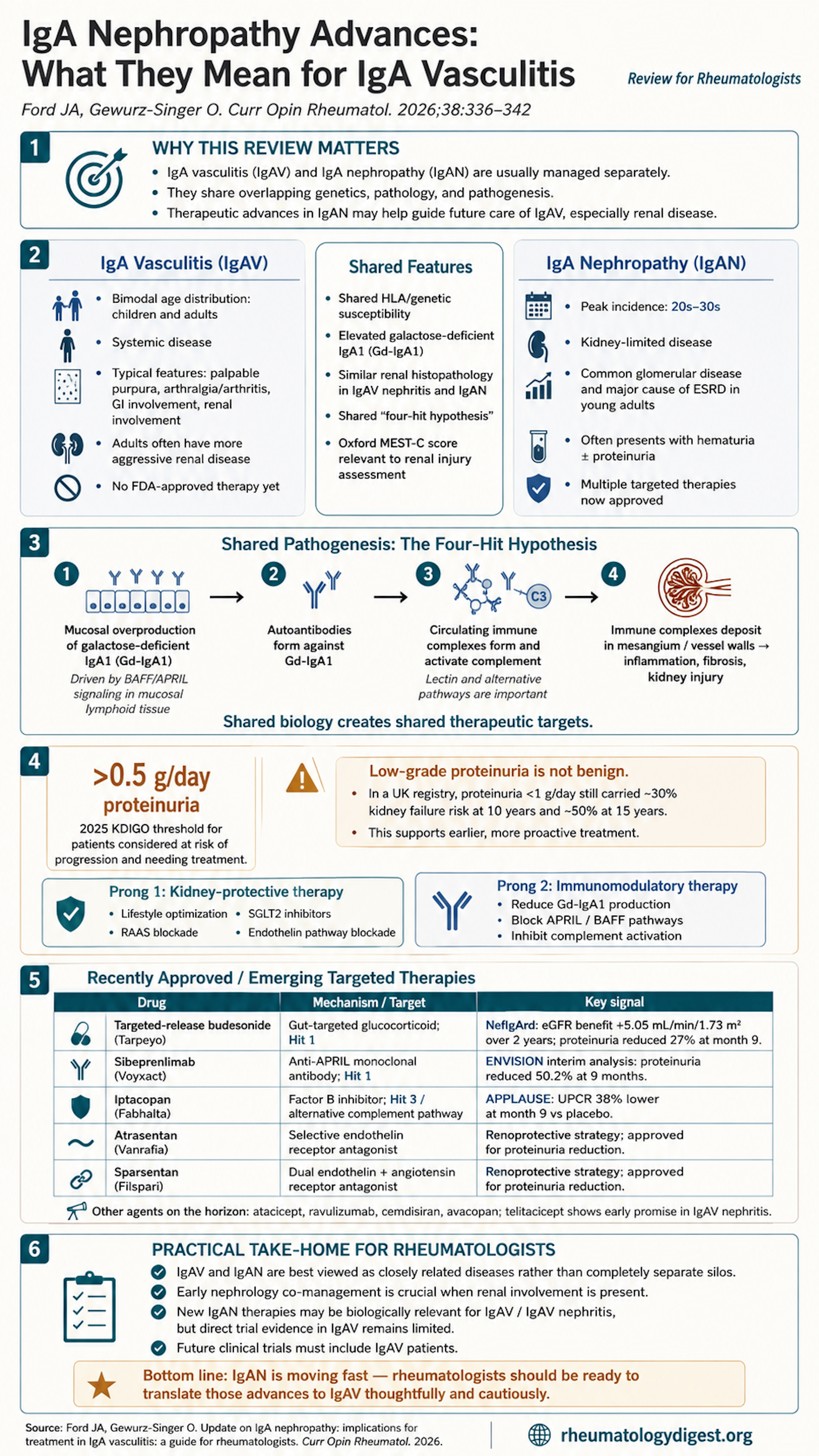
TL;DR: IgA vasculitis and IgA nephropathy look identical on renal biopsy and share their genetics and pathogenesis, but only one of them has approved drugs — and the immediate practice change is treating proteinuria above 0.5 g/day rather than 1.
The clinical problem
Two diseases with indistinguishable renal biopsies, shared genetics, and shared pathogenesis — one of which has five newly approved targeted therapies, and one of which has none.
IgA vasculitis (IgAV) — formerly Henoch–Schönlein purpura — is a systemic immune-mediated small-vessel vasculitis defined by a clinical tetrad: palpable purpura, arthralgia/arthritis (often lower-extremity), gastrointestinal involvement (abdominal pain through to haemorrhage or intussusception), and renal involvement. The nephritis is an immune complex-mediated glomerulonephritis spanning microscopic haematuria with minimal proteinuria to nephrotic-range proteinuria with elevated creatinine and hypertension. Urologic, neurologic and respiratory manifestations (including alveolar haemorrhage) are reported but rare.
The adult–paediatric split matters enormously. IgAV is the commonest systemic vasculitis of childhood, and paediatric disease is generally mild and self-limited. Adults comprise only 10–30% of cases but have a more aggressive course with more severe renal involvement — in a French cohort of 260 adult IgAV patients, 30% had renal failure at presentation. The rheumatologist’s IgAV population is therefore selected for exactly the phenotype the paediatric literature does not describe.
IgA nephropathy (IgAN) is the most commonly diagnosed glomerular disease in the United States (incidence ~1 per 100,000) and a leading cause of ESRD in young adults. Presentations range from recurrent gross haematuria (classically synpharyngitic, with upper respiratory infection) through microscopic haematuria ± proteinuria, to progressive renal dysfunction, nephrotic syndrome or RPGN.
The therapeutic asymmetry is the review’s animating grievance:
| IgA vasculitis | IgA nephropathy | |
|---|---|---|
| FDA-approved therapies | None | Five (two haemodynamic, three immunomodulatory) |
| Randomised controlled trials | Sparse; RIGA (rituximab + GC vs GC alone, NCT05329090) active in adults | Numerous, multiple positive phase III |
| Current management | Glucocorticoids; limited data for steroid-sparing DMARDs. Rituximab and mycophenolate mofetil show efficacy in small studies; cyclophosphamide has not shown convincing benefit | Two-pronged, guideline-directed, rapidly evolving |
Compounding this, IgAV patients have been systematically excluded from every IgAN trial — so the disease with the more severe systemic phenotype has been locked out of the therapeutic revolution occurring in its histopathological twin.
Are IgAV and IgAN the same disease?
The review assembles four independent lines of evidence:
1. Genetics. GWAS have identified shared HLA susceptibility in the HLA-DRB1 region, and a recent cross-phenotype GWAS identified 21 shared risk loci between IgAN and IgAV.
2. Histopathology — the strongest argument. Renal involvement in IgAV (IgAV with nephritis, IgAVN) is histopathologically indistinguishable from IgAN. Mesangial IgA deposition was described in both entities as early as 1969. The histological range in both spans mesangial proliferation → focal and segmental proliferation → severe crescentic glomerulonephritis.
3. Shared scoring systems. The Oxford MEST-C classification (M = mesangial hypercellularity, E = endocapillary hypercellularity, S = segmental glomerulosclerosis, T = tubular atrophy/interstitial fibrosis, C = crescents) correlates with kidney outcomes in IgAN and has been evaluated in IgAVN cohorts.
4. Shared serology. Serum galactose-deficient IgA1 (Gd-IgA1) is significantly elevated in both IgAN and IgAVN versus healthy controls.
A revealing asymmetry in the biopsy findings, and a plausible explanation. IgAVN tends to show more acute lesions (E and C), whereas IgAN shows more chronic changes (S and T). The authors’ interpretation is elegant: this likely reflects differential timing of diagnosis rather than differential biology. IgAVN announces itself through purpura, arthritis and abdominal pain; IgAN, being renal-limited, is silent until it is found. By the time IgAN is biopsied, chronicity has accrued. The lesion is the same; the clock is different.
The unresolved question the review is honest about: why do Gd-IgA1 immune complexes show tropism for renal mesangium in IgAN, while in IgAV they deposit less selectively across skin, joints, gut and kidney? This is not explained. The authors note that some have wondered whether patients with IgAN may have had unrecognised vasculitis in the past.
The four-hit hypothesis — the therapeutic map
This is the conceptual core of the review, because each hit is a druggable node.
| Hit | Event | Evidence in IgAVN specifically |
|---|---|---|
| Hit 1 | Overproduction of aberrantly glycosylated galactose-deficient IgA1 (Gd-IgA1) in mucosa-associated lymphoid tissue (MALT) of GI and upper respiratory tracts. In genetically predisposed individuals, an environmental stimulus at the mucosal surface upregulates BAFF and APRIL, promoting B-cell class switching to Gd-IgA1-producing B cells | Serum Gd-IgA1 significantly higher than controls in both IgAN and IgAVN |
| Hit 2 | Formation of autoantibodies against Gd-IgA1 (IgG and IgA class) | Serum anti-Gd-IgA1 levels correlate with disease activity in IgAVN |
| Hit 3 | Assembly of anti-Gd-IgA1–containing circulating immune complexes, mediated by complement | Lectin and alternative pathway markers elevated in IgAVN; skin and kidney biopsies show C3 and C5–C9 deposition |
| Hit 4 | Deposition of immune complexes in vessel walls and renal mesangium, activating mesangial cells and downstream inflammatory and fibrotic pathways | The point of divergence: selective mesangial deposition in IgAN, multi-organ in IgAV |
Why this framework matters clinically. It reframes IgAV from “a vasculitis we treat with steroids” to a mucosal B-cell/complement disease with four sequential, individually targetable steps. Every drug discussed below maps onto a specific hit — which is precisely what makes the extrapolation argument to IgAV coherent rather than merely hopeful.
Note also that Hit 1 originates at the mucosal surface, which explains both the classic synpharyngitic presentation and why a gut-targeted glucocorticoid became the first approved therapy.
The new treatment paradigm in IgAN — driven by two findings
Finding 1: “Low-risk” proteinuria is not low-risk.
The old paradigm — proteinuria <1 g/day means monitor off therapy — has collapsed under the weight of outcome data:
- UK National Registry of Rare Kidney Diseases (2,299 adults, 140 children with IgAN): patients with proteinuria <1 g/day had a 30% chance of kidney failure at 10 years and 50% at 15 years.
- Chinese cohort (n = 1,530): any proteinuria above normal (>0.3 g/day) conferred worse renal prognosis.
A 50% probability of kidney failure at 15 years in patients previously categorised as low-risk is a striking figure and the single most important number in the review. It should reset expectations for anyone who has been reassuring an IgAVN patient with 0.7 g/day of proteinuria.
Finding 2: These patients benefit from immunosuppression. The TESTING trial showed that glucocorticoids in patients with proteinuria improved renal outcomes versus no glucocorticoids.
Consequence — the KDIGO 2025 guideline (which explicitly covers both IgAN and IgAV) lowered the treatment threshold: patients with proteinuria >0.5 g/day are now defined as at risk of progressive kidney failure and in need of treatment.
The recommended two-pronged approach:
Prong 1 — Renal protective strategies (reduce nephron loss): lifestyle modification, RAAS blockade, SGLT2 inhibitors, and endothelin-1 receptor antagonists. Already standard of care in IgAVN and typically initiated by nephrology co-managers.
Prong 2 — Immunomodulation: target inflammation, prevent IgA immune complex formation and mesangial deposition. This is the prong the rheumatologist owns, and the one where the IgAN pipeline is most relevant.
The approved drugs — mapped to the four hits
| Drug (brand) | Mechanism | Hit targeted | Dosing |
|---|---|---|---|
| Atrasentan (Vanrafia) | Endothelin receptor antagonist | — (haemodynamic) | 0.75 mg daily PO |
| Sparsentan (Filspari) | Dual endothelin + angiotensin II receptor antagonist | — (haemodynamic) | 200–400 mg daily PO |
| Targeted-release budesonide (Tarpeyo/Nefecon) | Gut-targeted glucocorticoid | Hit 1 | 16 mg daily PO |
| Sibeprenlimab (Voyxact) | Anti-APRIL monoclonal antibody | Hit 1 | 400 mg SC every 4 weeks |
| Iptacopan (Fabhalta) | Complement factor B inhibitor | Hit 3 | 200 mg twice daily PO |
Hit 1 agents — reducing Gd-IgA1 production
Targeted-release budesonide (Nefecon). A slow-release oral glucocorticoid acting locally at the MALT to reduce Gd-IgA1 production — an elegant application of the mucosal-origin hypothesis. Accelerated FDA approval December 2021; full approval 2023 after the phase III NefIgArd trial.
- 364 patients with primary IgAN, all optimised on RAAS blockade, randomised to Nefecon 16 mg/day or placebo for 9 months, followed for 12 months.
- Primary outcome — time-weighted average eGFR over 2 years — significantly favoured Nefecon (difference 5.05 mL/min/1.73 m²).
- 27% proteinuria reduction versus placebo at month 9; 48% reduction at month 12, three months after stopping.
- Commonest adverse events: peripheral oedema and muscle spasms.
The persistence — and deepening — of the proteinuria effect after drug cessation is mechanistically interesting: it suggests the intervention modifies upstream Gd-IgA1 production rather than merely suppressing downstream inflammation.
Sibeprenlimab — anti-APRIL monoclonal antibody, FDA-approved November 2025. Interim analysis of the first 320 patients in the phase III ENVISION/VISIONARY programme: 400 mg SC every 4 weeks produced a 50.2% reduction in proteinuria versus a 21% increase on placebo at 9 months, with a similar safety profile between groups.
Atacicept — dual APRIL/BAFF inhibitor. Phase II ORIGIN: 35% proteinuria reduction versus placebo at 9 months; acceptable safety in open-label extension to 96 weeks. Phase III ORIGIN III has completed recruitment (NCT04716231).
Hit 3 agents — complement inhibition
Iptacopan — oral factor B inhibitor blocking the alternative pathway. Accelerated FDA approval August 2024. Pivotal APPLAUSE trial: 222 patients with IgAN and UPCR >1 g/g, iptacopan 200 mg BD versus placebo. At month 9, UPCR was 38% lower with iptacopan (P < 0.001), with a tolerable side-effect profile, no unexpected safety findings and no increase in infection risk.
Terminal pathway agents (investigational):
- Ravulizumab (long-acting anti-C5 mAb): phase II in 43 IgAN patients — proteinuria −41.9% versus −16.8% with placebo (30.1% treatment effect, P = 0.005). Phase III enrolling (NCT04201262).
- Cemdisiran (siRNA suppressing hepatic C5 production, SC): phase II in 31 patients showed improvement over placebo.
- Avacopan (C5a receptor inhibitor, repurposed from ANCA-associated vasculitis): modest efficacy in a small open-label pilot of 10 IgAN patients.
The avacopan datum is worth flagging for rheumatologists specifically — it is the one agent in this list already familiar from our own formulary, and the pilot evidence is correspondingly thin.
The emerging IgAV evidence — small, but pointing somewhere
This is the section rheumatologists should read most closely, because it is the only direct evidence in the review.
Telitacicept (dual BAFF/APRIL inhibitor — the same TACI-Fc fusion protein appearing in the SLE literature):
- Adult IgAVN: a small single-arm study of 13 patients showed significant proteinuria improvement. A phase II trial is underway.
- Paediatric IgAV/IgAVN: a retrospective study of 24 children treated with telitacicept versus 30 matched controls on conventional therapy showed significantly reduced proteinuria at 36 weeks.
- The finding that should interest rheumatologists most: in that paediatric study, five patients with extrarenal IgAV only reported improvement in gastrointestinal, skin and joint symptoms.
That last observation opens a genuinely new question: could BAFF/APRIL blockade treat non-renal IgAV? If Hit 1 is a shared upstream event and the extrarenal manifestations derive from the same immune complexes, there is no mechanistic reason it should not. The evidence for this is currently five uncontrolled paediatric patients, which is precisely the level at which enthusiasm should be tempered — but the hypothesis is coherent and testable.
Additional retrospective studies have reported efficacy of anti-APRIL/BAFF agents in IgAVN, including in refractory childhood disease.
Key takeaways
Conceptual
- IgAV and IgAN are plausibly one disease with two distributions of immune complex deposition. Shared HLA-DRB1 susceptibility, 21 shared GWAS risk loci, identical renal histopathology, shared Gd-IgA1 elevation, and a common four-hit pathogenic model. The organising question is no longer whether they are related but why deposition is mesangium-restricted in one and multi-organ in the other — which remains unanswered.
- The biopsy differences (acute E/C lesions in IgAVN, chronic S/T in IgAN) probably reflect diagnostic timing, not distinct biology. Extrarenal symptoms bring IgAVN patients to attention earlier.
- The four-hit model is a therapeutic map, not just a pathogenesis diagram. Hit 1 → gut-targeted budesonide, anti-APRIL, anti-BAFF. Hit 3 → factor B, C5, C5aR inhibition. Knowing which hit a drug targets is how you reason about extrapolation.
Practical — what changes on Monday
- Lower your proteinuria threshold. KDIGO 2025 — which explicitly covers IgAVN — defines >0.5 g/day as the threshold for at-risk disease requiring treatment. The old “<1 g/day is low risk” framing is refuted: 30% kidney failure at 10 years and 50% at 15 years in that supposedly low-risk group.
- Both prongs, not one. Renal protection (RAAS blockade, SGLT2 inhibitors, endothelin receptor antagonists, lifestyle) plus immunomodulation. Prong 1 is usually nephrology-led, but the rheumatologist should confirm it is happening.
- Co-manage IgAVN with nephrology deliberately. The therapeutic developments relevant to your IgAV patient’s kidneys are occurring in a different specialty’s literature and are being incorporated into a nephrology guideline.
- Current IgAV therapeutics remain thin: glucocorticoids first, with rituximab and mycophenolate mofetil supported by small studies, cyclophosphamide unconvincing, and no FDA-approved agent. The phase III RIGA trial (NCT05329090) is the one to watch.
- Off-label use of IgAN agents in IgAV is mechanistically defensible but evidentially unsupported. No IgAN trial included IgAV patients. The strongest direct evidence is 13 adults and 24 children on telitacicept, uncontrolled or retrospectively controlled.
Where the evidence is weakest — read the numbers carefully
- Almost every efficacy figure quoted is a proteinuria reduction, not a hard renal outcome. Only NefIgArd reports an eGFR-based primary endpoint (2-year time-weighted average, difference 5.05 mL/min/1.73 m²). Proteinuria is a validated surrogate in IgAN, but a 38% or 50% proteinuria reduction is not the same claim as preserved kidney function, and several of these approvals are accelerated on precisely that basis.
- Several pivotal results are interim analyses. Sibeprenlimab’s approval rests on an interim analysis of the first 320 patients at 9 months. Iptacopan’s APPLAUSE data are at month 9 in 222 patients.
- Sample sizes for the terminal complement agents are very small — 43 (ravulizumab), 31 (cemdisiran), 10 open-label (avacopan).
- The IgAV evidence is essentially anecdotal. Thirteen adults, single-arm. Twenty-four children, retrospective with matched controls. Five patients with extrarenal-only disease reporting symptomatic improvement. These generate hypotheses; they do not support practice change.
- Complement inhibition carries meningococcal risk that this review does not discuss — a real consideration for any rheumatologist contemplating C5 blockade off-label, and one the ANCA and PNH literatures address in detail.
- The review does not address cost, access, or how any of this applies outside high-income settings — relevant given that IgAN is disproportionately prevalent in Asian populations.
Final take
The argument this review makes is simple and, on the evidence assembled, hard to resist: we have two conditions with shared genetic risk loci, shared serology, shared pathogenesis and renal biopsies that cannot be told apart — and we have systematically excluded one of them from every trial that transformed the other. The consequence is that a rheumatologist managing adult IgAVN, a population in which 30% present with renal failure, has no approved therapy, while a nephrologist managing the same histological lesion now has five.
For practice today, the actionable changes are modest but real: treat at 0.5 g/day rather than 1 g/day, ensure both prongs of the KDIGO approach are running, and co-manage closely. For the field, the actionable change is larger — stop excluding IgAV patients from IgAN trials, and let the shared pathogenesis be tested rather than merely asserted. Until that happens, extrapolation from IgAN to IgAV remains what the authors carefully call it: mechanistically plausible, and unproven.