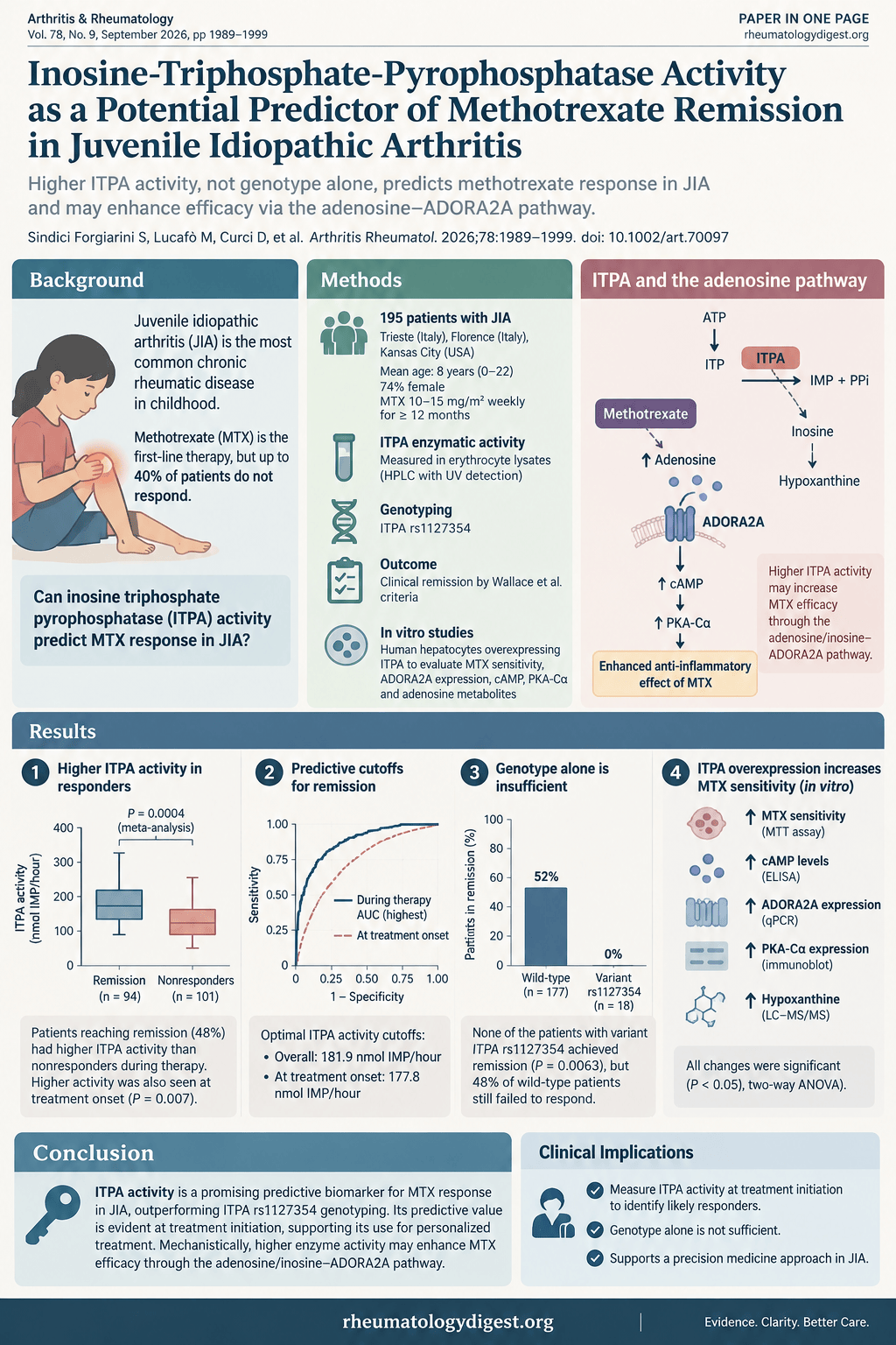
TL;DR: Erythrocyte ITPA activity measured before the first dose of methotrexate separates children with JIA who will reach remission from those who won’t, and predicts better than genotyping the ITPA variant does — but at roughly 60% sensitivity it shifts your prior rather than making the decision for you.
The problem worth solving
Methotrexate is first-line therapy in JIA, cheap, safe and effective enough that it remains the default after decades. And yet up to 40% of patients do not respond, which means disease progression and permanent joint damage accumulating during the months it takes to establish that fact.
There is no established way to know in advance. That is the gap this study is aiming at: a biomarker measurable at treatment initiation that separates likely responders from likely non-responders, so the latter could be escalated to a biologic earlier rather than after a failed trial.
Why ITPA is a plausible candidate
The reasoning runs through methotrexate’s actual anti-inflammatory mechanism, which is not the folate-antagonist story most of us learned first.
As a folate analogue, MTX inhibits DHFR and thymidylate synthetase, interfering with de novo purine and pyrimidine synthesis. But folic acid supplementation does not abolish the anti-inflammatory effect, which tells you something else is going on. The most widely accepted mechanism is adenosine accumulation, driven mainly by inhibition of AICAR transformylase (ATIC). Extracellular adenosine binds adenosine receptors — principally ADORA2A — and downstream signalling inhibits NF-κB, the key regulator of inflammatory gene transcription.
Two observations support this rather elegantly: the adenosine receptor antagonists caffeine and theophylline nullify the MTX effect in a rat adjuvant arthritis model, and patients with RA who consume large quantities of caffeine show reduced responsiveness to MTX.
Where ITPA fits. Inosine triphosphate pyrophosphatase preserves nucleotide homeostasis by hydrolysing non-canonical purine triphosphates — (deoxy)inosine triphosphate and (deoxy)xanthine triphosphate — into their monophosphate derivatives. Inosine monophosphate (IMP) can be converted to AMP, and AMP onward to adenosine. So higher ITPA activity plausibly feeds the adenosine pool that MTX depends on — a direct molecular link between an enzyme you can measure in erythrocytes and the pathway that makes the drug work.
The study
Children meeting ILAR criteria for JIA, either MTX-naïve or off MTX for at least 10 months, enrolled from three paediatric rheumatology centres: IRCCS Burlo Garofolo (Trieste), Meyer Children’s Hospital (Florence) and Children’s Mercy (Kansas City, Missouri). Patients with systemic JIA or on concomitant biologics were excluded.
195 patients, treated between 2000 and 2022 — 134 in Italy, 61 in the US. Mean age 8 years (range 0–22), 74% female, 90% White. Weekly MTX at 10–15 mg/m², oral or subcutaneous. Samples at baseline (up to 4 weeks pre-treatment), 6 months, then every 3 months, minimum 12 months follow-up.
Remission was defined stringently, by Wallace criteria: six unbroken months of clinically inactive disease on MTX, with that period starting inside the first year of therapy. Clinically inactive disease meant no active joints, no systemic symptoms, no uveitis, ESR and/or CRP normal, physician global assessment at best score, and morning stiffness under 15 minutes — with no other drugs during that period, including biologics, NSAIDs, and oral or intra-articular steroids. That is a demanding endpoint, and worth holding in mind when reading the 48% remission rate.
ITPA activity was measured in erythrocyte lysates by HPLC with UV detection, quantifying IMP as the hydrolysis product. ITPA rs1127354 — a non-synonymous variant known to reduce enzyme activity — was genotyped separately.
The main finding
93 of 195 patients (48%) achieved clinical remission. Sex was not associated with either remission or ITPA activity.
Patients who reached remission had higher ITPA activity than those who did not, at both time frames:
| Remission | No remission | |
|---|---|---|
| All time points during therapy | 212.1 ± 90.8 nmol IMP/h | 169.6 ± 81.5 |
| At therapy onset | 232.4 ± 92.0 nmol IMP/h | 183.7 ± 84.3 |
The association held after adjustment for clinical site — logistic regression P = 0.0006 and meta-analysis P = 0.0004 for all measurements during treatment; P = 0.003 and P = 0.007 respectively at therapy onset.
The clinically important part is that second row. ITPA activity predicted response when measured before the first dose, which the authors state is the first demonstration of measuring ITPA activity pre-treatment for this purpose. A biomarker that only works once you have been on the drug for six months is of limited use; one that works at initiation could change the first decision you make.
ITPA activity did not change significantly between therapy onset and subsequent time points (203.3 ± 91.7 vs 193.3 ± 98.5 nmol IMP/h, P = 0.38), nor within the patients who achieved remission (P = 0.65) — so this looks like a stable patient characteristic rather than something MTX itself modifies.
A site effect worth noting. ITPA activity differed significantly between centres (Kruskal-Wallis P = 4.62 × 10⁻⁵ across all measurements), with Trieste patients showing lower activity than both Florence and Kansas City. The authors attribute this to population stratification and differing genetic backgrounds — consistent with previous multicentre pharmacogenetic studies — and adjusted for site as a covariate throughout. A likelihood ratio test showed no significant effect of site on remission itself.
The cutoffs, and how much weight they bear
ROC analysis produced two thresholds:
| Cutoff | Sensitivity | Specificity | Remission above cutoff | Remission below cutoff | |
|---|---|---|---|---|---|
| All time points | 181.9 nmol IMP/h | 60% | 63% | 60% (56/94) | 37% (37/101) |
| At therapy onset | 177.8 nmol IMP/h | 67% | 58% | 55% (28/51) | 30% (14/46) |
Read those honestly. A test with 60% sensitivity and 63% specificity is not a decision rule. Being above the onset cutoff takes your probability of remission from a 48% base rate to 55%; being below takes it to 30%. That is a real and reproducible shift — the difference between roughly one in three and roughly one in two — but it is a nudge, not a verdict. The authors say as much: integration with other biomarkers or clinical factors may be necessary for clinical usefulness.
Enzyme activity beats genotype
This is the part I found most interesting, and it is a useful general lesson about pharmacogenetics.
ITPA rs1127354 was genotyped in 129 patients; 8 carried the variant, with a minor allele frequency of 3.9% consistent with White population data. As expected, activity was much lower in variant carriers (58.2 ± 40.1 vs 186.0 ± 73.6 nmol IMP/h, P = 6.2 × 10⁻⁶) — the P32T mutation alters enzyme structure and reduces activity.
None of the 8 variant carriers achieved remission (Fisher’s exact P = 0.0063, corrected OR 18.45, 95% CI 1.04–326.82). That looks striking until you read the confidence interval, which spans from barely-above-1 to 327 — eight patients cannot support a precise estimate, and this finding needs replication before it means anything at the bedside.
The more useful observation is the converse: 48% of wild-type patients also failed to respond, and among wild-type patients, higher ITPA activity was still associated with greater likelihood of remission (P = 0.044). Genotype therefore captures only part of the signal. Measuring the enzyme captures more than inferring it from one SNP — which makes sense, since activity integrates every genetic and non-genetic influence on the protein, not just one variant.
The mechanism, tested in vitro
To probe why, the authors used immortalised human hepatocytes (IHH) stably transfected either with an ITPA-containing plasmid or an empty vector.
- MTX sensitivity. After 72 hours, ITPA-overexpressing cells were markedly more sensitive to MTX — IC50 7.2 ± 5.39 nM versus 35.6 ± 72.4 nM in mock-transfected cells.
- cAMP. No difference at 24 hours, but intracellular cAMP was higher in ITPA cells at 48 hours at both MTX concentrations tested (25 nM and 200 nM — chosen to approximate levels reached in vivo after oral dosing).
- PKA-Cα. No change at 24 or 48 hours; higher expression in ITPA cells at 72 hours.
- NF-κB. No difference at 24 or 48 hours, but at 72 hours mock cells showed higher phosphorylated p65 than ITPA cells at the same MTX concentrations — consistent with less NF-κB activation where ITPA is abundant.
- ADORA2A expression did not differ between the lines at baseline (P = 0.76), so the effect is on ligand availability and downstream signalling, not receptor abundance.
The metabolite data are more equivocal and the authors handle them carefully. Extracellular adenosine was actually higher in mock cells at 200 nM MTX, while hypoxanthine was higher in ITPA cells under all conditions including untreated. Their explanation: because the stop solution was added 30 minutes before the end of incubation, adenosine already released by ITPA cells could have been metabolised onward — to inosine via ecto-adenosine deaminase, then hypoxanthine via purine nucleoside phosphorylase — implying faster and greater adenosine release followed by degradation, rather than less release.
They also raise a second possibility that does not require adenosine at all: the elevated hypoxanthine might come from inosine derived from IMP, and inosine is itself anti-inflammatory — it suppresses IL-8 in human epithelial cells, decreases TNF-α in vitro and in vivo, and acts both independently and through ADORA2A. In a murine pleurisy model, adenosine and inosine at sub-effective doses were synergistically anti-inflammatory. So inosine and adenosine may act together in high-ITPA cells.
An alternative mechanism that has nothing to do with adenosine
Worth registering, because it would change what the biomarker means. ITPA activity might influence MTX response through competition between ITP and MTX for folylpolyglutamate synthetase.
Low ITPA activity → ITP accumulates → interferes with MTX polyglutamate formation → less intracellular active MTX metabolite → reduced efficacy. High ITPA activity clears ITP, removes the competitive inhibition, and permits optimal polyglutamation.
The authors call this a plausible biochemical explanation for the same clinical observations, and — importantly — they did not measure MTX polyglutamate levels, which they list as a limitation in establishing the mechanistic basis. So the adenosine story is supported but not established over this alternative.
How much to believe
The authors’ acknowledged limitations:
- Relatively small sample and a single-ancestry cohort used to calculate the cutoffs, with no replication cohort currently available. The 90%-White composition limits generalisability, and larger numbers would give more reliable thresholds.
- Sensitivity and specificity are moderate, as above — the authors present the cutoffs as a first step toward clinical translation rather than a finished tool.
- MTX polyglutamates were not measured, leaving the mechanistic link incomplete.
- In vitro constraints: limited biological replicates, and a hepatocyte line that does not represent the cell types involved in JIA pathophysiology. The stated aim was to isolate ITPA’s effect on MTX response in a controlled system, but confirming this in a disease-relevant cell model is still needed.
- Single-time-point adenosine measurement limits understanding of its dynamic metabolism; selective ADORA2A antagonists would be needed to confirm receptor involvement.
One further thing the paper does not address, which I would want to see. In Table 1, the mean age at MTX start was 6 years in the remission group and 10 years in the non-remission group, and the JIA subtype distribution also differs between them — persistent oligoarticular disease (the subtype with the better prognosis) made up 53 of 93 remitters versus 44 of 102 non-remitters, while polyarticular RF-negative disease ran the other way, 28 versus 37. The primary analysis is adjusted for clinical site, but I could not find adjustment for age or JIA subtype. Both are plausibly associated with remission independent of ITPA, so some of the observed association could reflect case mix rather than enzyme activity. This does not undo the finding — the effect was consistent across three sites and present at baseline — but it is an unaddressed confounder in a study whose whole purpose is prediction.
What this could change
- The concept is the advance, not yet the test. A pre-treatment assay that stratifies MTX responders in JIA would be genuinely useful, and this is the first demonstration that ITPA activity carries that signal before the first dose. That is worth knowing even though the test is not ready.
- If it replicates, the proposed use is escalation, not exclusion. The authors suggest patients with low ITPA activity — at baseline or during treatment — might be assigned to more aggressive therapy, such as early initiation of a biologic. Note the framing: low activity would argue for adding something, not for withholding methotrexate.
- Measured enzyme activity beat inferred genotype. For any pharmacogenetic marker where a functional assay exists, this is a reminder that the assay may carry more information than the SNP — 48% of wild-type patients here still failed to respond.
- Do not use the cutoffs clinically yet. Single cohort, no replication, one ancestry, moderate discrimination. The numbers are a starting point for the next study, not a threshold for the next clinic.
- A small aside with immediate applicability: the caffeine data. If MTX works substantially through adenosine receptor signalling, and adenosine receptor antagonists nullify it in animal models while high caffeine intake associates with reduced MTX responsiveness in RA, that is at least worth asking about in a patient responding poorly.