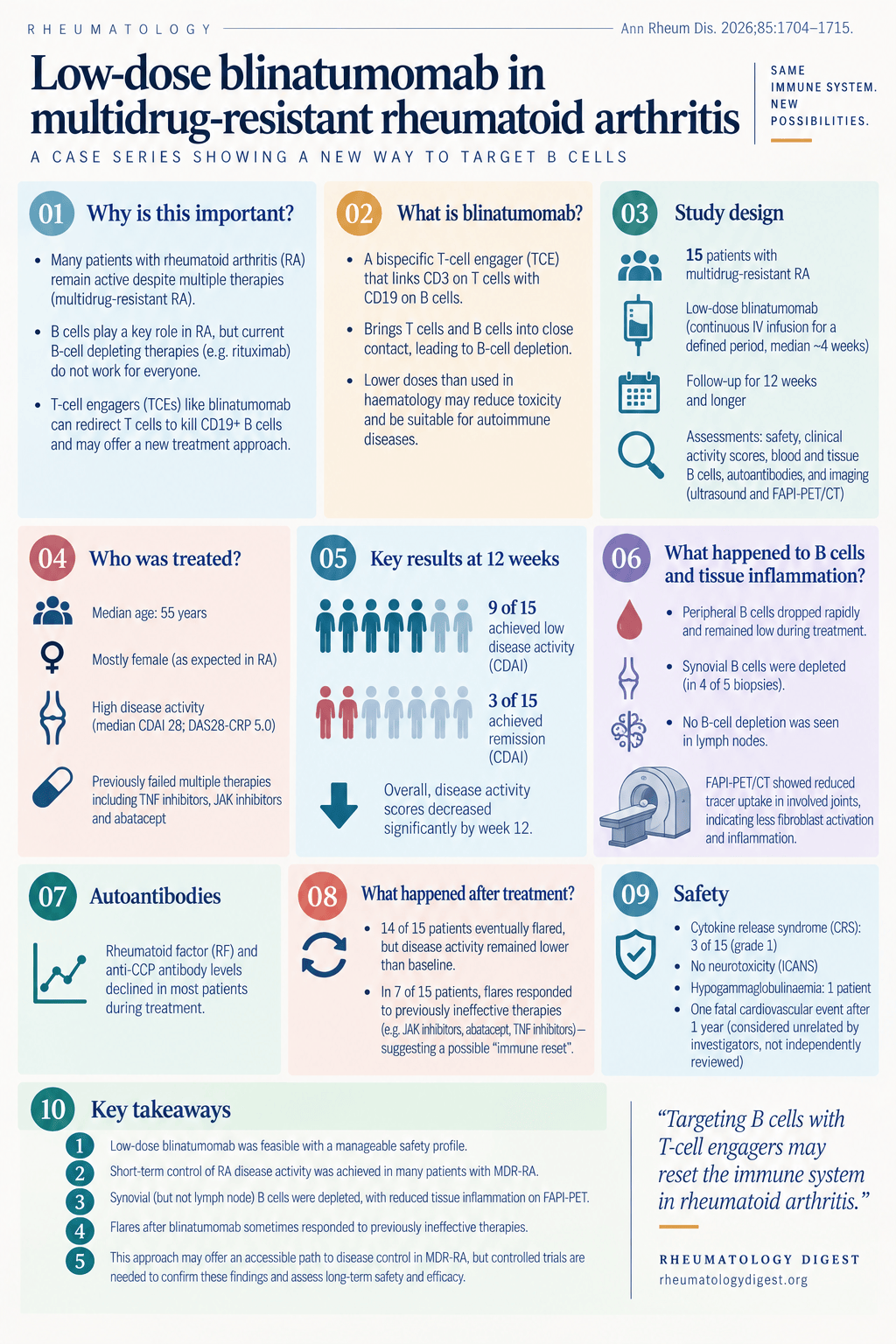
TL;DR: At roughly a tenth of the haematology dose, blinatumomab produced ACR20 in all 15 patients with multidrug-resistant RA and cleared synovial B cells — but 14 of 15 flared, and the interesting finding is that seven then responded for over three months to drugs that had previously failed them.
The question behind the dose
Patients with RA who have failed multiple DMARDs are a genuine therapeutic dead end, and deep B-cell depletion — via CAR-T cells or T-cell engagers — has become the most talked-about way out. The implicit goal in most of that work is immune reset: eradicate the pathogenic B-cell clone completely and let the immune system rebuild.
Blinatumomab is the first approved T-cell engager — two linked single-chain variable fragments targeting CD3 on T cells and CD19 on B cells. At 54 kDa it is a third the size of an immunoglobulin, and its 1–2 hour half-life means it needs continuous infusion but is rapidly reversible, which matters when things go wrong. In B-cell ALL it can eradicate minimal residual disease.
The trade-off is that the doses which maximise depletion in haematology also carry real risks of cytokine release syndrome and neurotoxicity. So this study asks a narrower and arguably more practical question: can a deliberately low dose modify RA meaningfully, even if it never achieves full immune reset?
That framing is the point. The authors are explicit that therapeutic concepts beyond full reset are gaining interest, on the view that different depths of B-cell depletion may serve different clinical purposes.
What was done
A named-patient (compassionate use) case series, not a trial. Fifteen patients treated at Erlangen between April 2023 and November 2024 — selected from 38 considered, with 11 excluded for not meeting severity criteria, 8 for not meeting resistance criteria, and 4 who declined consent.
Entry required RA by 2010 ACR/EULAR criteria, evidence of B-cell involvement (RF, ACPA, antimodified peptide antibodies, ANA, or B cells in the synovial membrane), DAS28-CRP above 3.2, and failure of methotrexate plus at least three different b/tsDMARDs.
This was a genuinely refractory cohort: median age 55 (range 27–64), 60% female, median disease duration 13 years (range 3–33), median DAS28-CRP 5.03, CDAI 28, SDAI 27. Two-thirds (10 of 15) were refractory to rituximab.
The dose is the story. Per cycle, 36–38.5 μg was given by continuous infusion over 96 hours (9 μg/day), with a one-week treatment-free interval before cycle two. Five patients had the dose raised to 28 μg/day in cycle two; four received a third cycle. Cumulative dose ranged 77 to 189 μg.
For comparison, approved haematology dosing runs up to 784 μg per cycle on a 28-day continuous infusion. So this is roughly a tenth of the exposure, delivered over a quarter of the time, with breaks — a low-dose, interrupted protocol by design.
Glucocorticoids were tapered to ≤10 mg before starting; methotrexate was continued in six patients; all other DMARDs stopped at least three weeks beforehand. Patients received 16 mg dexamethasone as premedication before each infusion cycle — remember this, because it becomes the single most important confounder.
Assessment was unusually thorough for a series this size: clinical scores, flow cytometry, serum immunoglobulins and cytokines, autoantibodies, musculoskeletal ultrasound, sequential synovial biopsy in 5 patients, lymph node biopsy in 4, and FAPI-PET/CT to image synovial fibroblast activation.
Safety
This is the most reassuring part of the paper.
- Three cases of CRS, all grade 1. One resolved with metamizole; two needed a single dose of tocilizumab. Notably, CRS was not clearly dose-linked — two events occurred on day one of the first infusion, one during a dose increase at cycle three.
- No ICANS. No neurotoxicity at all, which the authors contrast with the haematology experience and read as evidence that TCEs behave more favourably in autoimmune disease than in oncology.
- Five mild infections in 4 of 15 (26.6%) — herpes labialis, SARS-CoV-2, a UTI, dysuria that resolved without antibiotics, and a periodontitis. No severe infections.
- One patient developed mild hypogammaglobulinaemia at week 3 (IgG 449 mg/dL), needed no immunoglobulin replacement, and normalised by week 6.
- Immunoglobulins overall remained stable, as did free kappa and lambda light chains.
The death needs stating plainly. Patient #9 — pre-existing coronary artery disease, 40 pack-year smoking history — suffered a fatal cardiovascular event one year after treatment. The treating investigators categorised it as unrelated, but there was no independent adjudication and no autopsy. The authors are appropriately careful here: although most cardiovascular events after TCE therapy in oncology occur within the first weeks to months, delayed events are reported, and they conclude that long-term cardiovascular monitoring is warranted in high-risk patients receiving T-cell redirecting therapy. Given a single-arm series of 15 patients, this event cannot be attributed or dismissed with any confidence.
B-cell depletion: blood versus tissue
T-cell engagement caused a transient fall in circulating CD3⁺ cells without altering the CD4/CD8 ratio, alongside a transient rise in acute-phase proteins and proinflammatory cytokines during the first infusion.
In peripheral blood, depletion was fast but short-lived. Three patients (#5, #7, #10) were already depleted from prior rituximab. Of the rest, two depleted after the first infusion, five after the second, two after the third — and three patients (#2, #4, #11) never achieved full peripheral depletion at all. Median time to B-cell recovery was just 21 days (range 7–51).
The tissue findings are where this gets interesting:
| Compartment | Finding |
|---|---|
| Peripheral blood | Fast, short B-cell aplasia; median recovery 21 days; 3 of 15 never fully depleted |
| Synovium (5 biopsies) | CD20⁺ B cells fell to zero in 4 of 5 |
| Inguinal lymph node (4 biopsies) | B cells reduced but not depleted |
No significant change in lymph node plasma cells, T cells or macrophages — and one of the four biopsied patients showed a post-treatment increase in lymph-node CD138⁺ plasma cells, consistent with the known resistance of long-lived plasma cells to CD19-directed therapy.
That dissociation — synovium cleared, lymph node not — is the mechanistic hinge of the paper, and it is also where the authors are most careful about over-reading their own data (see caveats).
On autoantibodies: by month 3, RF fell by an average 62.1%, aMCV by 54%, anti-CCP2 by 45% — then rose again between months 3 and 6, coinciding with returning disease activity.
Short-term clinical response
Rapid, and in all 15 patients. By week 12:
| Measure | Baseline (median) | Week 12 (median) |
|---|---|---|
| Tender joint count | 7 | 2 |
| Swollen joint count | 5 | 1 |
| Patient VAS (0–100) | 70 | 35 |
| DAS28-CRP | 5.03 | 2.4 |
| CDAI | 28 | 8 |
Response rates: all 15 reached at least ACR20; 11 of 15 (73%) reached ACR50; 8 of 15 (53%) reached ACR70. By CDAI, 3 of 15 achieved remission (≤2.8) and 9 of 15 reached low disease activity or better (≤10) — only patient #15 remained in high disease activity. Five of 15 (33%) attained DAS28-CRP remission by month 3.
In a cohort where everyone had failed methotrexate plus three or more targeted agents and two-thirds had failed rituximab, an ACR50 rate of 73% is a striking number. Hold that alongside the fact that it is uncontrolled and steroid-premedicated.
The finding that may matter more than the response rate
14 of 15 patients flared. Only patient #11 remained in drug-free remission (at 9 months). Disease activity rose again after an average of 5 months (range 1–18).
So on the immune-reset criterion, this failed. But what happened next is the paper’s most provocative observation.
Because the cohort was so refractory, reintroduced drugs were often ones patients had already failed — six were rechallenged with the same agent, three switched within the same class. And:
- At month 3 after reintroduction, disease activity improved on abatacept, on JAK inhibitors, and to a lesser extent on TNF inhibitors — despite 2 of 5 abatacept patients, 4 of 5 JAK-inhibitor patients, and all TNF-inhibitor patients having previously failed that exact mechanism.
- Overall, 9 of 15 were retreated with a previously-received or same-class drug, and 7 of 15 achieved a clinical response lasting more than three months.
The authors’ framing is that post-blinatumomab flares represented a de-escalated phenotype — still active disease, but no longer multidrug-resistant. If that holds up, it reframes what low-dose TCE therapy is for: not a cure, but a way to restore the effectiveness of the drug armamentarium you already have.
Three competing explanations, and the authors name all three:
- Reduced synovial fibroblast activation. Synovial B-cell depletion — even without lymph node depletion — may disrupt local B-cell–fibroblast interactions, consistent with the FAPI-PET findings. The authors explicitly flag this as a hypothesis for the later tissue and serological findings, not an explanation for the early clinical response, which preceded all tissue assessment.
- T-cell exhaustion induced by the TCE could add to the effect of cytokine inhibitors.
- A drug holiday. The period off the failed agent during blinatumomab therapy may simply allow pharmacokinetic resensitisation — which would reverse secondary loss of response without any immunological reset at all.
That third explanation is the deflationary one, and it is important that the authors note secondary loss of response was the most frequent reason these drugs had failed in the first place. Pharmacological resensitisation applies primarily to secondary, not primary, failure.
Tissue and imaging
- Ultrasound showed a clear reduction in synovitis after treatment.
- FAPI-PET/CT in 5 patients showed overall reduced tracer uptake — i.e. less synovial fibroblast activation. The one exception was patient #1, whose FAPI uptake increased at 3 months, at a point when they had already flared.
- Lower post-treatment FAPI scores, particularly in the hands, were associated with a longer duration of initial clinical response before further therapy was needed. That is the closest thing here to a candidate imaging biomarker of durability — from five patients, so treat it as a hypothesis.
How much to believe
The authors’ limitations section is unusually forthright, and there are several things worth adding to it.
Their own caveats:
- This is an uncontrolled, exploratory case series. No control arm, no randomisation, n=15.
- The dexamethasone confounder. Patients received 16 mg dexamethasone per infusion cycle, and the authors accept that early improvement within the first two weeks may partly reflect this. Their counter-argument is reasonable: sustained benefit over months, responsiveness to previously failed therapies, and tissue-level changes are unlikely to be explained by dexamethasone alone. But the early response genuinely cannot be separated from steroid effect.
- Biopsy timing was heterogeneous — lymph node 29–97 days, synovium 29–242 days, including one biopsy taken after three cycles at day 242. This may sample active B-cell repopulation rather than residual post-treatment burden, and precludes definitive comparison between synovial and lymph node depletion — which is the paper’s headline mechanistic claim. The authors say so themselves, and also note that lymphocyte infiltration, retention, residence and egress may simply differ between the two tissues.
- Subgroup analyses were not randomised. The dose and prior-rituximab comparisons reflected physician discretion and may be subject to responder-selection bias.
- Within-class switching cannot be separated from the resensitisation claim.
Additional considerations:
- Selection is substantial. Fifteen of 38 patients considered were treated, and the eligibility filter was applied by specialised rheumatologists. This is the most refractory tail of an already-selected referral population — which cuts both ways: it makes the response rate more impressive and the generalisability weaker.
- Ultrasound readers were not blinded to clinical data (they were blinded to the FAPI images), and evaluation deliberately targeted the most clinically affected joint — a selection the authors themselves flag as a potential bias.
- The FAPI scoring system is not standardised. The 3-point SUV-max scale was, by the authors’ own description, intended to capture overall trends rather than serve as a validated measure.
- The fatal cardiovascular event was not independently adjudicated, as above.
- Patients and the public were not involved in design, conduct, reporting or dissemination — worth noting for a compassionate-use series in a severely affected population.
- Competing interests are extensive across the senior authorship (consulting, advisory and speaking relationships with a long list of manufacturers), though no commercial sponsor was involved in the study and funding came from DFG and academic sources.
What this changes
- Nothing in practice today. This is 15 patients, uncontrolled, on compassionate use. It is a signal-generating study, and the authors say as much — causal inference and generalisability require controlled trials.
- The safety signal is the most transportable finding. Grade 1 CRS only, no ICANS, no severe infections, stable immunoglobulins with no replacement needed. If low-dose TCE protocols in autoimmune disease consistently look like this, it changes the risk calculus for trialling them at all — particularly against the CAR-T comparison, which requires lymphodepletion.
- Partial depletion may be enough to be useful. The most quotable conclusion is that total eradication of the pathogenic B-cell clone may not be the only route to durable clinical benefit. Synovium cleared while lymph node did not, and patients still improved substantially for months.
- “Resensitisation” is the hypothesis worth testing properly. Seven of 15 responding for over three months to drugs that had already failed them is either a genuinely new therapeutic concept or an artefact of drug holidays and within-class switching. A controlled trial could separate those; this design cannot.
- Short peripheral B-cell aplasia (median 21 days) with clear synovial depletion is a reminder that circulating B-cell counts are a poor proxy for tissue effect — relevant whenever you use peripheral CD19 counts to judge whether a B-cell-directed therapy has “worked”.
- If you follow this field, the open question is dose. Patients with higher cumulative exposure trended toward better responses, and the authors suggest higher doses plus a subcutaneous formulation to extend half-life might deepen tissue depletion. That is the next experiment — balanced against the safety profile that makes the low-dose approach attractive in the first place.
For the broader mechanistic context on this drug class in autoimmune disease, see T-cell engagers in rheumatology — which made the argument that rheumatology has been under-dosing these agents relative to oncology. This series is a direct test of the opposite proposition, and the answer appears to be “partly, and usefully, but not enough for reset.”