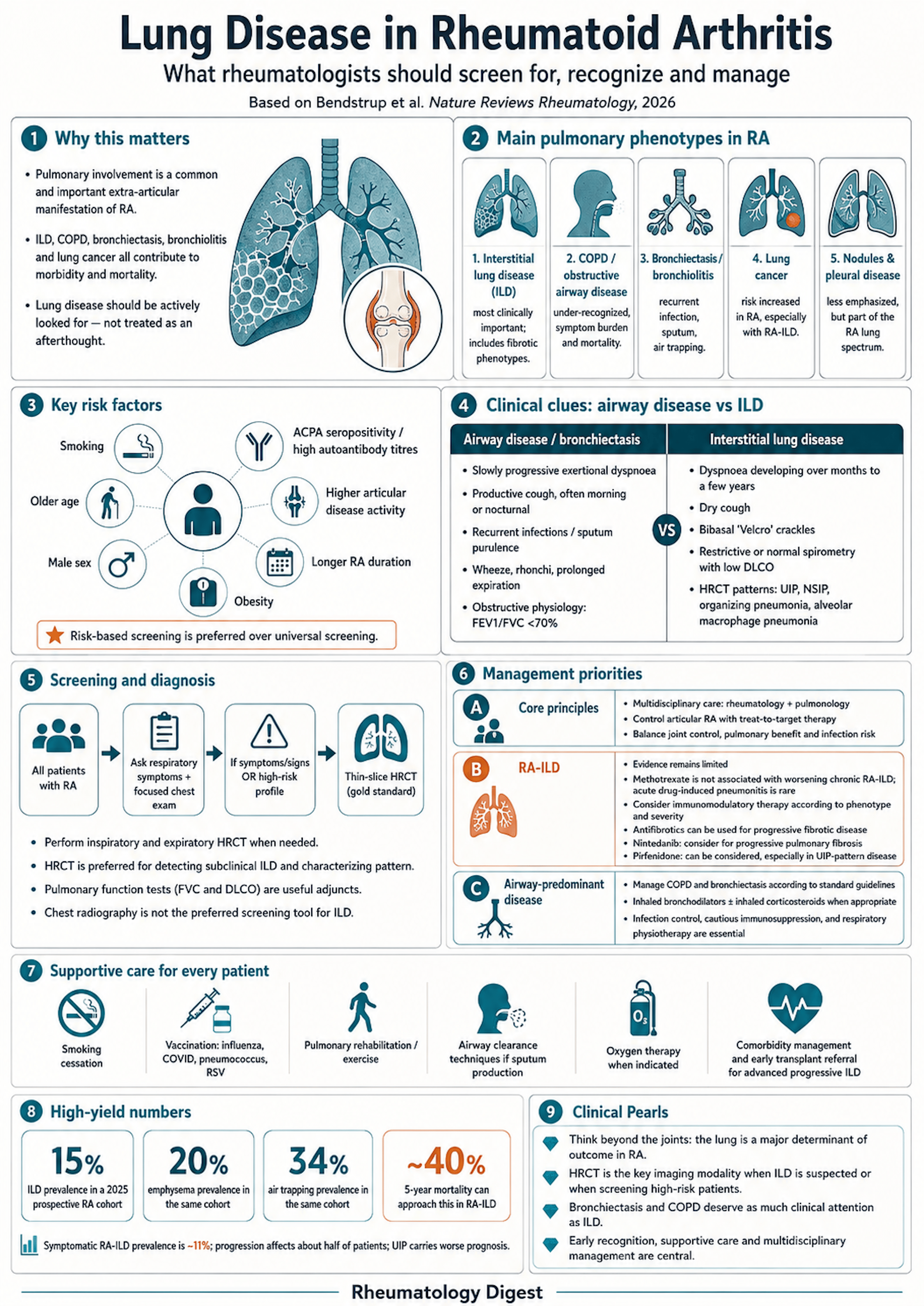
TL;DR: Pulmonary involvement is a leading contributor to reduced survival in RA, but only ILD has attracted screening guidance — COPD, bronchiectasis and bronchiolitis are common, carry their own mortality, and complicate every immunosuppression decision you make.
Why the lung deserves more attention than it gets
RA affects roughly 1% of the global population, predominantly women in their fifth and sixth decades. The systemic inflammation that drives it produces extra-articular manifestations, and pulmonary involvement together with cardiac comorbidity now represents the primary contributor to reduced survival in RA.
The respiratory diseases most commonly associated with RA are the airway diseases — COPD and bronchiectasis — alongside interstitial lung disease. Patients are also at increased risk of lung cancer, rheumatoid nodules and pleural disease. These conditions are distinct but frequently overlap: COPD, bronchiectasis, bronchiolitis, ILD and lung cancer often co-occur in one patient, which matters because those distinctions drive treatment choice and follow-up.
Smoking is the thread running through all of it. It is a well-established risk factor for ACPA-positive RA, and that risk factor is shared with COPD, ILD and lung cancer — all of which are more prevalent in RA than in the general population. A direct causal relationship between smoking and these pulmonary conditions has not, however, been definitively established.
Prevalence figures remain imprecise, largely because screening methods, diagnostic approaches and cohort characteristics differ so widely between studies. A 2025 multicentre prospective study evaluated 258 patients within 1–10 years of RA diagnosis using HRCT and lung function, and reported prevalences of ILD 15%, emphysema 20% and air trapping 34%.
The asymmetry the review is arguing against: screening for cardiac disease is now an integral part of RA management, and ILD screening is moving in the same direction — the 2025 ERS–EULAR guidelines recommend basing the decision to screen with HRCT on risk factors including age, male sex, high autoantibody titres and smoking history. Far less emphasis has been placed on screening for COPD and bronchiectasis, despite both being common in RA and associated with poor prognosis. Both carry considerable morbidity, symptom burden, reduced quality of life and decreased survival, and the authors argue they warrant the same rigour as RA-ILD.
Obstructive airway disease
Pathophysiology. Several factors contribute: small-airway inflammation and remodelling, with lymphoid follicle formation in terminal bronchioles, mucus plugging and concentric fibrosis limiting airflow and causing air trapping. The mucosal lining of the airway is one plausible location for tolerance to break down and for citrullination to occur, and local airway inflammation can predate joint disease. Microbial colonisation and infection — including Pseudomonas species — are common in RA and amplify neutrophilic inflammation, accelerating airway damage. Susceptibility to infection becomes particularly relevant when immunosuppression is required for the joints. Smoking synergises with ACPA seropositivity to drive both RA and COPD, and seropositive patients are at increased risk of COPD compared with seronegative patients.
COPD. In RA, an obstructive phenotype driven mainly by small-airway pathology, bronchiolitis and bronchiectasis — often related to current or previous smoking — is common yet under-recognised. One prospective cohort found obstructive functional limitation on spirometry in 20.7% of patients with RA, while HRCT identified airway abnormalities (bronchial wall thickening, bronchiectasis, mosaic attenuation) in 61% of those patients. Structural airway disease substantially exceeds what lung function testing detects — a point worth holding onto when spirometry comes back normal. Small-airway limitations were common in that study and associated with symptoms and impaired quality of life. A population-based meta-analysis put the 5-year cumulative probability of COPD at 7.4% in RA versus 6.0% in matched individuals without RA.
Asthma, and a bidirectional relationship. Studies over the past 10–15 years show a significant and potentially bidirectional association between asthma, chronic rhinitis, nasal polyps and RA. A meta-analysis of six cohort and 14 case–control studies reported a pooled HR of 1.42 (95% CI 1.18–1.70) for RA among patients with asthma, and Mendelian randomisation supports a causal link, suggesting adult-onset asthma might increase RA risk (OR 1.018). In a Korean population-wide study, RA was associated with asthma (OR 2.32, 95% CI 1.51–3.57), allergic rhinitis and sinusitis. Asthma can be more severe in people with RA, with higher in-hospital mortality and costs. The summary position: asthma seems to increase the risk of RA, while RA is linked to greater prevalence and severity of asthma.
Treatment. Control the underlying disease and minimise airway exposure to noxious particles, mainly tobacco smoke. Inhaled bronchodilators — long-acting β2 agonists or long-acting muscarinic antagonists — with or without inhaled corticosteroids are reasonable in symptomatic and asthma-like obstruction. Fixed, non-reversible obstruction in constrictive bronchiolitis responds only modestly to inhalation therapy. Macrolides could reduce exacerbations and cough in bronchiolitis or bronchiectasis phenotypes, and selected patients might benefit, balanced against antimicrobial stewardship. Airway clearance techniques, vaccination, smoking cessation and pulmonary rehabilitation all matter.
Evidence for managing airway-predominant lung disease in RA with csDMARDs or bDMARDs is sparse, and no specific treatment recommendations exist for RA-associated COPD beyond standard COPD guidelines. Immunomodulatory agents used in RA could confer elevated risk of pulmonary infection. One intriguing signal: in a nationwide Danish observational cohort, the risk of hospitalised acute COPD exacerbation or death over a 6-month period was reduced in methotrexate users compared with matched non-users, and the authors suggest the potential role of methotrexate in mitigating COPD-like patterns warrants further study.
Bronchiolitis
A rare manifestation that can present pathologically as constrictive bronchiolitis obliterans, follicular (cellular) bronchiolitis, or less commonly diffuse panbronchiolitis.
Bronchiolitis obliterans often presents with severe irreversible airway obstruction and is clinically indistinguishable from COPD — progressive breathlessness, cough (sometimes productive), rarely wheezing, reduced exercise capacity. Patients are typically far more symptomatic than the imaging would lead you to expect. The main feature distinguishing it from COPD is the lack of, or very subtle, smoking history, and often a protracted disease course. There is a female preponderance, and almost all patients develop it after the RA diagnosis rather than before. Drug-induced bronchiolitis has been reported with penicillamine, sulfasalazine and gold salts.
Physiology and imaging. Pulmonary function testing reveals severe obstruction with a low FEV1/FVC ratio, typically preserved or only mildly reduced DLCO, and increased residual volume on body plethysmography due to air trapping. Inspiratory HRCT can show bronchial wall thickening, bronchiectasis, centrilobular emphysema and nodular opacities with mosaic attenuation — especially in follicular bronchiolitis — while expiratory scans show pronounced air trapping. Ground-glass opacities, mild reticulation or even honeycombing in the lower lobes can coexist.
Diagnosis and treatment. Bronchoscopy with lavage can rule out infection; the differential white cell count usually shows a neutrophilic, non-specific inflammatory pattern. Transbronchial biopsies are rarely diagnostic, and surgical lung biopsy is rarely performed because the complication risk usually outweighs the benefit of a confident diagnosis. In most patients the clinical, physiological and radiological picture suffices. In the largest case series — 41 patients with RA-associated bronchiolitis obliterans — all-cause mortality was 27% over 5 years. Treatment literature is limited: supportive bronchodilators and inhaled corticosteroids are often initiated, responses to systemic glucocorticoids, cyclophosphamide, azathioprine and etanercept are variable and must be weighed against infection risk, and some case series report stabilisation or improvement with macrolides. Many patients do not respond, and lung transplantation evaluation should be considered.
Bronchiectasis
The coexistence of RA and bronchiectasis is conceptualised as bronchiectasis rheumatoid overlap syndrome (BROS).
HRCT is the diagnostic modality of choice, typically revealing dilated bronchi with thickened walls. On cross-sectional images the bronchial diameter usually exceeds that of the accompanying pulmonary artery — the signet ring sign. This differs from traction bronchiectasis, which sits selectively within areas of fibrotic parenchyma, and bronchiectasis in RA is often seen in the outer third of the lung, which helps distinguish it.
Epidemiology, and a large gap between symptoms and imaging. RA confers an elevated risk of incident bronchiectasis (adjusted HR 2.12), with higher prevalence in seropositive than seronegative disease. Prevalence estimates vary enormously by ascertainment: symptomatic bronchiectasis is found in roughly 2.7–3.1% of patients with RA, whereas systematic HRCT identifies it in 22.6–24.9%. Crucially, BROS is independently associated with severely impaired outcomes including increased mortality — a mortality risk nearly double that of idiopathic bronchiectasis.
Which comes first? Two competing models:
- The airway-first model proposes that underlying genetic risk — heterozygosity for CFTR variants — or innate immune defects such as undetectable circulating mannose binding lectin cause chronic bronchial inflammation and subsequent infection. The resulting antigenic stimulation, potentially involving externalisation of citrullinated peptides via neutrophil extracellular traps, triggers systemic autoimmunity and ACPA generation, leading to RA. This is historically supported by cohort studies in which bronchiectasis symptoms preceded RA in a high proportion of patients (83.3–93.8%).
- The RA-first model is supported by more recent longitudinal data showing RA antedates incident bronchiectasis, suggesting systemic rheumatic inflammation drives the pulmonary disease. The risk is substantially more pronounced in seropositive RA (adjusted HR 2.34) than seronegative (HR 1.56), illustrating the central role of high-grade rheumatic inflammation.
Other risk factors include older age, longer RA duration and specific HLA variants.
The treatment bind. DMARDs — conventional and biologic — along with glucocorticoids form the backbone of RA treatment and increase susceptibility to recurrent lower respiratory tract infection, including mycobacteria. This often leads clinicians to avoid bDMARDs in patients with known bronchiectasis. Treatment must therefore target the underlying bronchiolar and articular inflammation without compromising antibacterial defences. A promising strategy under exploration is inhibition of dipeptidyl peptidase 1 by the neutrophil elastase inhibitor brensocatib, which reduces the frequency of pulmonary exacerbations and could address the chronic inflammatory component of BROS while reducing the burden of recurrent infection.
Interstitial lung disease
Pathophysiology, and the case for the lung as the origin of RA
RA-ILD results from the interaction of genetic susceptibility, environmental factors and immune dysregulation. The MUC5B promoter variant rs35705950 is the only well-established genetic variant significantly associated with increased RA-ILD risk, and its pathogenic effect is restricted to patients with a UIP pattern on HRCT, with an effect comparable in magnitude and direction to that reported in idiopathic pulmonary fibrosis. Beyond genetics, premature cellular senescence has emerged as a pivotal mechanism.
Smoking induces epithelial injury and impairs repair in the lung parenchyma, facilitating fibrotic remodelling. High RA disease activity is a risk factor for both incident and prevalent ILD.
The mucosal origins hypothesis is the conceptually important part. Two lines of evidence support early pulmonary involvement in the development of RA itself: environmental exposures such as cigarette smoke and silica are important risk factors for ACPA-positive RA; and elevated ACPA levels are detected in sputum samples from patients with RA, including those with early-stage disease. Together these suggest citrullination within the pulmonary mucosa could initiate local autoimmunity and thereby initiate RA pathogenesis. Temporal analyses reinforce this — ILD can precede or co-occur with the RA diagnosis in 17–48% of cases. The specific contribution of locally produced ACPAs remains to be fully elucidated, but the lung is a candidate primary site rather than merely a target organ.
Epidemiology and prognosis
The UIP pattern predominates, with a pooled prevalence of approximately 46% in patients with RA-ILD — a marked contrast with ILD associated with other connective tissue diseases, where other radiological patterns are more common.
Combined pulmonary fibrosis and emphysema — emphysema apically, fibrosis in the lower zones — has a prevalence of up to 48% in RA-ILD. It is physiologically characterised by preserved lung volumes with significantly reduced DLCO, and carries a high risk of pulmonary hypertension and lung cancer. This is a pattern where normal-looking spirometry actively misleads.
Short telomeres are a recognised risk factor, with more pronounced shortening in patients with a UIP pattern. They are strongly associated with increased ILD prevalence, greater severity, accelerated lung function decline and worse outcomes — and, importantly, patients with short telomeres can experience reduced survival when exposed to immunosuppression.
Other robust risk factors: older age at RA onset, male sex, seropositivity with elevated ACPA titres, increased disease activity and prolonged disease duration. Modifiable factors include smoking and obesity.
| Ascertainment method | ILD prevalence in RA |
|---|---|
| Symptomatic ILD (2023 systematic review and meta-analysis) | ~11% |
| Systematic HRCT screening of asymptomatic patients (three studies) | 11–21% |
| Clinically indicated HRCT, counting interstitial lung abnormalities | 44% |
That last row deserves emphasis: a retrospective analysis of 293 patients undergoing clinically indicated chest HRCT identified interstitial lung abnormalities — subtle findings not meeting diagnostic criteria for ILD but now considered preclinical ILD — in 44%, underscoring the considerable burden of subclinical involvement.
Prognosis. Disease progression affects approximately half of patients with RA-ILD, whether symptomatic or asymptomatic, and is associated with a mortality risk exceeding both the general population and patients with RA without ILD. Survival after diagnosis is variable, with median times ranging between 2.6 and 8 years and a 5-year mortality approaching 40%. The UIP pattern consistently correlates with worse outcomes across cohorts, and a meta-analysis of 1,256 patients highlighted pulmonary function metrics and extent of lung involvement as prognostic indicators.
Screening
Screening means evaluating patients without respiratory symptoms — distinct from diagnostic assessment, which is triggered by clinical manifestations and for which chest HRCT is the gold standard. For screening, HRCT should be performed without contrast using a thin-slice technique (commonly <1 mm), with inspiratory and expiratory phases to detect air trapping.
The most characteristic HRCT patterns, in decreasing frequency: UIP, NSIP, organizing pneumonia, and alveolar macrophage pneumonia (formerly termed desquamative interstitial pneumonia). Drug-induced ILD should always be considered in the differential, particularly when organizing pneumonia is identified and when ILD develops within weeks to months of starting a new medication.
Screening is best conceptualised as two-tiered: a systematic assessment of respiratory symptoms and focused clinical examination in all patients with RA, with any relevant symptom or sign escalating to definitive diagnostic evaluation; then a more intensive surveillance strategy applied selectively to patients at elevated risk.
Both the 2025 ERS–EULAR and 2023 ACR–CHEST guidelines favour a risk-based rather than universal approach — but neither explicitly defines the criteria identifying high-risk populations, and neither endorses implementation of published risk-prediction models. A screening algorithm incorporating commonly assessed variables such as age at RA onset, sex and disease activity has shown potential for early detection. Both sets discourage chest radiography, invasive diagnostic procedures and chest ultrasonography for screening, designating HRCT as preferred. They diverge on pulmonary function tests: ERS–EULAR excluded FVC and DLCO as screening tools, whereas the ACR–CHEST panel advocated their use alongside HRCT.
Management
Current recommendations are largely extrapolated from RCTs focused on articular disease and from retrospective observational studies. They distinguish between immunomodulatory agents — primarily for articular control, with potential ILD benefit — and antifibrotics targeting fibrosis, used alone or in combination based on individualised risk–benefit assessment.
On methotrexate — the myth that needed correcting. Methotrexate has long been suspected of worsening pre-existing chronic RA-ILD. Although it can rarely induce acute or subacute drug-induced pneumonitis, retrospective, registry-based and case–control studies have consistently demonstrated that methotrexate is not associated with an increased risk of worsening chronic RA-ILD. On the contrary, patients treated with methotrexate show better survival, longer time to ILD development and fewer respiratory hospitalisations than patients not receiving it — though the authors note some bias cannot be excluded.
Management aims to slow progression, prevent acute exacerbations and improve survival. Controlling articular disease with a treat-to-target strategy using csDMARDs (methotrexate, sulfasalazine, leflunomide) and bDMARDs (rituximab, abatacept) per EULAR guidelines is essential, as these agents might also benefit the ILD. Mycophenolate mofetil is often used in practice but does not treat articular disease, and the evidence for its use in RA-ILD is very weak.
Two scenarios warrant ILD-targeted therapy: at diagnosis in the case of advanced disease (moderate or severe ILD), and on documented progression meeting established criteria for progressive pulmonary fibrosis.
| ACR–CHEST 2023 | ERS–EULAR 2025 | |
|---|---|---|
| Criteria for starting ILD-targeted therapy | Not specified | Apply the inclusion criteria of RCTs in CTD-ILD |
| PFTs in screening | Advocated alongside HRCT | Excluded as screening tools |
| Antifibrotics | Nintedanib and pirfenidone both considered second-line options | Nintedanib for progressive pulmonary fibrosis; pirfenidone for a UIP pattern |
The evidence problem, stated plainly. Over the past 25 years numerous RCTs have established evidence-based treatments for RA — yet no prospective RCT has shown the efficacy of these treatments in RA-ILD. Only one phase II trial has been conducted specifically in RA-ILD: TRAIL1, comparing pirfenidone with placebo in 123 patients. It was terminated early due to slow recruitment during the COVID-19 pandemic and did not reach its primary endpoint of reducing disease progression — though FVC decline was attenuated in the total population and more so in the subgroup with a UIP pattern on HRCT.
The antifibrotic evidence is largely borrowed. In INBUILD and FIBRONEER-ILD, nintedanib and nerandomilast respectively slowed progression in progressive pulmonary fibrosis — but only 89 of 663 patients in INBUILD and 118 of 1,176 in FIBRONEER-ILD had RA-ILD.
For advanced progressive ILD despite maximal medical therapy, lung transplantation is viable, and early referral for evaluation is strongly recommended. Overall, current guidance rests on low-certainty evidence.
Lung cancer
RA confers a clinically meaningful excess risk of lung cancer that extends beyond that conferred by shared risk factors such as smoking. Large contemporary cohorts show a 50–60% higher incidence than in non-RA populations, with the highest risk concentrated in patients with RA-ILD. A 2024 US Veterans Health Administration cohort linked RA to a >50% increased risk overall, and RA-ILD to an approximately fourfold higher risk. Nationwide data from Asia and the USA support this, and suggest the elevated risk persists even among never-smokers, underscoring RA-specific mechanisms.
Proposed mechanisms include chronic airway inflammation, citrullination, ectopic lymphoid structures within the lung, and cumulative exposure to infection. The presence of RA-ILD — comparably to idiopathic pulmonary fibrosis — increases risk probably via fibrotic remodelling, altered immune microenvironments and diagnostic surveillance effects. Whether methotrexate, other csDMARDs or bDMARDs contribute to lung cancer risk or recurrence has produced conflicting results.
Whether the elevated risk justifies dedicated lung cancer screening has not been established — but when screening for lung cancer, concomitant screening for ILD is warranted. Worsening cough, dyspnoea, thoracic pain, weight loss and fatigue should prompt timely referral, attention should be paid to the possibility of lung cancer whenever a chest CT is performed, and smoking cessation should be promoted.
On checkpoint inhibitors: pre-existing autoimmune rheumatic disease, including RA, does not preclude the benefit of ICIs, as it does not confer greater risk of mortality or of severe immune-related adverse events compared with matched controls. Patients with RA and lung cancer were more likely to have immunotherapy-related adverse events in a retrospective cohort, but this was mostly restricted to mild RA flares or other mild effects. Coordinated management across rheumatology, thoracic oncology and pulmonology is key.
Rheumatoid nodules
Pulmonary rheumatoid nodules are benign, often found incidentally and typically asymptomatic, though some patients present with cough, haemoptysis or pleuritic pain. They are more common in smokers and in those with severe, seropositive RA, and occur in approximately 3.5% of patients with RA.
The primary clinical challenge is differentiating them from lung cancer, since avid uptake can be seen on ¹⁸F-FDG-PET in both. Imaging features favouring benign nodules on CT include multiplicity, calcification, cavitation, smooth borders and peripheral subpleural location. Accurate diagnosis is essential given overlap with infectious and malignant processes, and sometimes requires histopathology — which shows a core of fibrinoid necrosis ringed by palisading histiocytes, with chronic inflammatory change around it.
Nodules generally do not require specific treatment unless symptomatic or complicated by infection or haemorrhage. Methotrexate and anti-TNF agents have been implicated in nodule development, so it is important to consider discontinuing these first before assessing impact. Subsequent treatment with hydroxychloroquine, rituximab or JAK inhibitors can be associated with stabilisation or regression, while surgical excision remains an option in refractory cases with diagnostic uncertainty.
Pleural disease
Common manifestations are pleurisy, pleural effusions and subpleural rheumatoid nodules. Pleural involvement is often subclinical and primarily identified on imaging; large effusions can present with cough, dyspnoea, chest pain and fever.
Reported prevalence varies widely — symptomatic pleural disease occurs in <5% of cases, some studies report frequencies as high as 20%, and post-mortem analyses show pleural effusions in about 50% of patients. The frequency of clinically apparent pleural disease is declining over time.
Effusions are more frequent in middle-aged men with high-titre seropositive disease, antinuclear antibodies and nodules. Pleural complications are often late manifestations but can precede joint symptoms. Effusions in RA are usually unilateral exudates and can co-occur with pericardial effusions. Macrophage-rich effusions are non-specific and could suggest other causes, whereas lymphocytic predominance can suggest RA; neutrophil-predominant effusions are usually infectious but can occasionally occur in RA.
Chronic or recurrent large effusions can produce pleural thickening and fibrosis. Thickening involves both parietal and visceral layers and can progress to fibrothorax, causing trapped lung and restrictive pathophysiology. Rarely it mimics malignancy, necessitating biopsy. Histology usually shows non-specific pleural inflammation with epithelioid and multinucleated giant cells dominating over mesothelial cells.
Subpleural nodules might cavitate and rupture into the pleural space, leading to pneumothorax, bronchopleural fistula, empyema and secondary infection; rarely, sterile empyematous effusions or pseudochylothorax follow rupture of necrobiotic nodules. Drug toxicity has been reported to cause accelerated nodulosis and recurrent pneumothoraces.
Management is guided by symptom burden and underlying pathology, but evidence-based guidance for symptomatic cases remains lacking. Asymptomatic effusions can be observed; symptomatic cases signal aggressive systemic disease and are treated with systemic glucocorticoids and DMARDs. Refractory or recurrent effusions might require repeated thoracentesis, pleurodesis or surgery — and certain DMARDs have been implicated in drug-induced pleural disease, making medication review necessary.
Distinguishing the three main entities at the bedside
| COPD | Bronchiectasis | ILD | |
|---|---|---|---|
| Symptoms | Increased dyspnoea over years, morning cough, sputum | Dyspnoea, cough, sputum purulence, recurrent infections | Asymptomatic, or increased dyspnoea over months to years, dry cough |
| Auscultation | Prolonged expiration and/or rhonchi | Normal, or rhonchi and/or crackles | Normal, or Velcro crackles |
| Spirometry | Irreversible obstruction, low FEV1, normal or low FVC, FEV1/FVC <70% | Obstruction with low FEV1, normal or low FVC, FEV1/FVC <70% | Normal or equally decreased FEV1 and FVC |
| Plethysmography and diffusion | High TLC and residual volume; decreased DLCO | Normal or high TLC and residual volume; normal or decreased DLCO | Normal or low TLC and residual volume; normal or decreased DLCO |
| HRCT | Emphysema | Bronchiectasis | UIP, NSIP, organizing pneumonia, alveolar macrophage pneumonia, or indeterminate |
| Treatment | LABA and/or LAMA and/or ICS | LABA and/or LAMA and/or antibiotics | Immunomodulation and/or antifibrotics |
| Follow-up | General practice or pulmonologist | General practice or pulmonologist | ILD centre, with pulmonology and rheumatology |
Two clinical discriminators worth committing to memory: digital clubbing suggests ILD but its absence does not exclude it, and DLCO declines early in ILD but also falls in advanced COPD with emphysema — so a reduced DLCO does not by itself point to the parenchyma.
Supportive care, which is where much of the benefit lives
Given how thin the disease-modifying evidence is, the adjunctive measures carry disproportionate weight: smoking cessation programmes; minimising environmental and occupational inhalational exposure; vaccination against influenza, COVID, pneumococcus and respiratory syncytial virus; Pneumocystis prophylaxis with co-trimoxazole for patients on high-dose glucocorticoids; pulmonary rehabilitation with endurance and resistance training; airway clearance techniques in patients producing sputum; long-term oxygen therapy for chronic hypoxaemia (≥15 h daily) with ambulatory and nocturnal oxygen as indicated; management of reflux, sleep apnoea, cardiac comorbidity and pulmonary hypertension; lung volume reduction in emphysema and early transplantation evaluation; attention to glucocorticoid-induced myopathy and osteopenia; mental health; and palliative care including advance care planning.
Where this leaves practice, and what is still missing
- Screen for ILD on the basis of risk, not universally — age, male sex, high autoantibody titres, smoking history and disease activity — using non-contrast thin-slice HRCT with expiratory phases. Do not use chest radiography or ultrasound to screen.
- Do not let normal spirometry reassure you. Structural airway disease on HRCT far exceeds what lung function detects, and combined fibrosis-with-emphysema preserves lung volumes while DLCO falls.
- Stop treating methotrexate as an ILD hazard in chronic disease. The consistent signal across registry and case–control data points the other way, though acute drug-induced pneumonitis remains a rare and separate entity.
- COPD and bronchiectasis deserve the same rigour as ILD. BROS carries nearly double the mortality of idiopathic bronchiectasis, and its infectious character genuinely constrains bDMARD use — a management problem that needs solving rather than avoiding.
- A UIP pattern is the prognostic dividing line in RA-ILD, and it is the pattern in which the MUC5B effect, telomere shortening and worse survival all concentrate.
- Consider bronchiolitis obliterans in the obstructed patient who never smoked — the disproportion between symptom burden and imaging, plus a preserved DLCO with air trapping, is the giveaway.
The honest summary is that research in RA-ILD has intensified over the past 5–10 years while COPD and bronchiectasis have received far less attention, and that high-quality RCT evidence remains limited across all of it. Future trials will need standardised inclusion criteria, harmonised outcomes (FVC decline, DLCO, imaging endpoints and patient-reported measures) and adequate power in well-characterised subgroups — and will have to reckon with competing risks of infection and malignancy. Lung ultrasonography is emerging as a possible cost-effective, non-invasive first filter for identifying who should proceed to HRCT. Whether early antifibrotic therapy before patients meet criteria for progressive pulmonary fibrosis becomes the standard for preventing irreversible damage is, for now, an untested but plausible proposition.