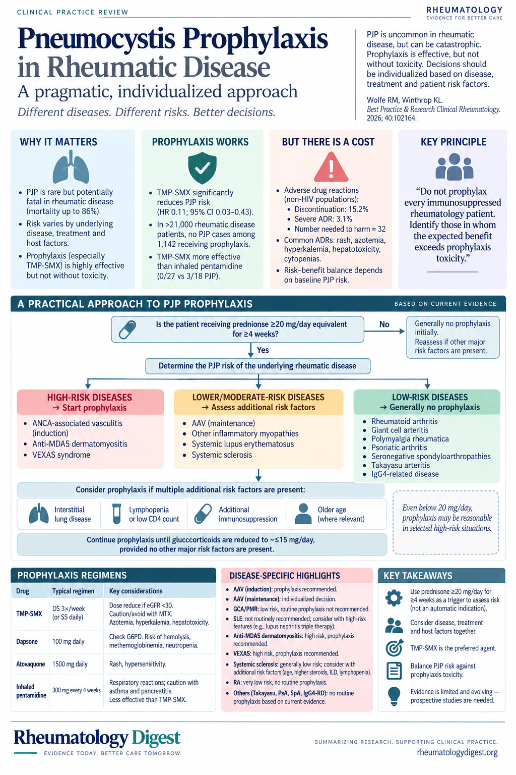
TL;DR: PJP prophylaxis in rheumatic disease cannot be reduced to a steroid threshold — the number needed to treat runs from about 4 in VEXAS to over 900 in rheumatoid arthritis, so the underlying disease decides at least as much as the dose.
Pneumocystis jirovecii pneumonia is uncommon in rheumatology, frequently fatal when it happens, and entirely preventable — which is precisely the combination that produces inconsistent practice. Evidence-based prophylaxis guidelines exist for HIV, transplantation and haematological malignancy. No such guidelines exist for rheumatic disease, despite steadily expanding exposure to potent immunosuppression.
The reasons are structural. Rheumatic diseases are individually uncommon, PJP incidence within any single disease is low, and absolute case numbers are therefore small. The diseases themselves are a heterogeneous group with distinct mechanisms, phenotypes and drug regimens. The data that exist are almost always retrospective and hard to generalise. So clinicians extrapolate or rely on judgement, and practice patterns vary substantially between providers. This review — from a rheumatologist and an infectious diseases specialist — tries to replace that with something defensible.
Prophylaxis works, and the mortality it prevents is high
Efficacy is not the uncertain part. In a large retrospective cohort of 818 patients, TMP-SMX significantly decreased PJP incidence with a hazard ratio of 0.11 (95% CI 0.03–0.43). In a larger database study of over 21,000 patients with rheumatic disease, there were no cases of PJP among the 1,142 patients who received prophylaxis. Randomised trials in ANCA-associated vasculitis, where prophylaxis is mandated per protocol, have demonstrated the same.
Agent choice matters. A retrospective study comparing TMP-SMX with inhaled pentamidine found 3 of 18 patients on inhaled pentamidine developed PJP, versus 0 of 27 on TMP-SMX — consistent with reduced efficacy of alternative agents seen in other immunocompromised populations. There are no other head-to-head studies among the second-line agents in rheumatic disease.
What raises the stakes is that mortality from PJP is higher in rheumatic disease than in HIV-infected individuals, with reported rates as high as 86%. This is not a nuisance infection.
The regimens. TMP-SMX is first line: one double-strength tablet three times weekly, one single-strength tablet daily, or one DS tablet daily. Cautions include dose reduction at eGFR <30 ml/min/1.73m², caution or avoidance with concurrent methotrexate, azotaemia, hyperkalaemia and hepatotoxicity. Dapsone (100 mg once daily, or 100 mg in two divided doses) requires caution at eGFR <30 or in G6PD deficiency, and carries methaemoglobinaemia, haemolytic anaemia, neutropenia and hypersensitivity. Atovaquone 1500 mg once daily can cause rash and hypersensitivity. Inhaled pentamidine 300 mg every four weeks causes respiratory reactions in up to 65% of patients, with caution in asthma and pancreatitis.
So why not give it to everyone?
Because the harms are real and the denominators are large.
A 2007 Cochrane review in non-HIV immunocompromised patients reported ADRs requiring discontinuation in 15.2% (40/263) and severe ADRs in 3.1% (9/288). That gives a number needed to harm of 32, and the conclusion that prophylaxis becomes favourable once PJP incidence exceeds 3.5% — a threshold most rheumatic diseases never approach.
Reported ADR rates in rheumatic disease vary considerably. One review of single-strength daily TMP-SMX showed an adverse event rate of 8.5%. In a study of 124 patients, overall ADR rates were 2.2% per patient-year, with TMP-SMX at 3% per person-year — and, notably, dapsone had the highest ADR rate at 9% per person-year. A large Japanese cohort reported an ADR rate of 21.2 per 100 person-years with serious ADRs at 1.2 per 100 person-years, giving an NNH of 131 if only serious events are counted, which is considerably more favourable. In a retrospective review of patients on rituximab with glucocorticoid exposure, the NNH for serious ADRs was 86.
Even so, patients and clinicians may reasonably regard rash, azotaemia, hyperkalaemia and laboratory abnormalities as unacceptable in the absence of meaningful PJP risk.
And the risk varies enormously by disease. A large population-based study from Hong Kong found an overall NNT in the 400s — but an NNT of 48 in AAV against 909 in rheumatoid arthritis. That spread is the whole argument of the paper.
Glucocorticoids: the most consistent risk factor, and why “30 mg” fails as a rule
Glucocorticoid exposure is the most consistent risk factor. The mechanism is likely multifactorial, reflecting both the steroid-induced decrease in circulating lymphocytes including CD4+ T cells and the impact on the alveolar response in the lung, though it remains incompletely understood. There is broad consensus that high-dose use increases risk; what counts as “high dose” has been far harder to pin down.
The most informative work comes from Park and colleagues, who studied patients on high-dose steroids defined as ≥30 mg/day prednisone equivalent for ≥4 weeks. The cohort spanned 17 rheumatological diseases — SLE 50.5%, Behçet’s 14.4%, the remaining third across 15 others. Over 1,747.4 person-years among 1,092 patients, 30 cases of PJP occurred: 29 in patients not receiving prophylaxis, and one after stopping prophylaxis for an ADR. The overall NNT was 52. Stratified by dose, it dropped to 32 at ≥60 mg/day and rose to 215 below 60 mg/day. Similarly, in a retrospective study of patients on rituximab plus steroids, the NNT for high-dose use was 20, against 250 for those not on high-dose steroids.
That dose-dependence led some authors to conclude that anyone on ≥30 mg/day for at least a month should receive prophylaxis. The review argues this has two flaws, and both are practical:
- It is not universally applicable across diseases. GCA often requires prolonged high-dose steroids, yet PJP rates stay low. High-dose steroid use may justify prophylaxis in some conditions, but the underlying disease has to be part of the calculation.
- The 30 mg cut-off misses patients. In the Chan study, prednisone ≥15 mg/day captured most cases. Even in Park’s own data, 40% of PJP cases occurred at doses between 15 and 30 mg/day.
When to stop remains debated. Risk is higher at higher doses and most cases occurred above 15 mg/day, but in both studies a handful of cases developed below that — and all of those patients had additional risk factors. The review’s position: once the dose is down to ≤15 mg/day, stopping prophylaxis is reasonable in the absence of other risk factors.
No single DMARD earns prophylaxis on its own
In a survey of rheumatologists, 68.8% reported that the therapeutic regimen was the most important factor in deciding whether to give prophylaxis, and real-world data confirm that treatment regimen is independently associated with receiving it. Case reports have implicated almost every drug in the rheumatological armamentarium.
But the evidence does not support singling out any agent. One study found immunosuppression was an independent risk factor in the first two weeks after starting steroids, with no differences between the immunosuppressive medications examined. More tellingly, individual drug risk appears to be modified by the presence of other risk factors: cyclophosphamide and rituximab may increase risk in ANCA vasculitis, yet the same drugs do not appear to carry the same risk in SLE and RA respectively. Immunosuppressant use belongs in the decision, but no single conventional or biologic DMARD warrants universal prophylaxis.
CD4 count and lymphopenia: useful, but no threshold exists
CD4+ T cells are central to immune clearance of Pneumocystis, and in HIV a count below 200 cells/mm³ is a well-established threshold. It would be convenient to extrapolate — but no such defining level is established in rheumatic disease.
Part of the gap is that CD4 testing is not part of routine rheumatological practice, so retrospective reviews have few data points in either direction. Patients with rheumatic disease who develop PJP do consistently show lower CD4 counts than matched counterparts, but the “at risk” threshold varies widely across studies, from 68 to over 300 cells/mm³ — and there are patients with equally low counts who never develop PJP.
Lymphopenia is likewise repeatedly established as a risk factor. One retrospective review showed both pre-treatment lymphopenia and lower lymphocyte counts during treatment were associated with increased risk; another found decreased counts at diagnosis and during admission even after controlling for steroid use.
The review’s conclusion for both markers is the same and worth holding onto: neither should be an indication for prophylaxis in isolation, but in patients with other established risk factors they help inform the risk-benefit assessment.
Interstitial lung disease and age
ILD is common in rheumatic disease and is treated with high-dose steroids, so its appearance as a risk factor is unsurprising. The susceptibility is likely driven by disruption of alveolar macrophage function — a primary cell in Pneumocystis clearance — along with active inflammation, compounded by the steroids used to treat it downregulating other cytokines important for clearance. This plausibly explains the higher PJP rates in diseases like AAV and anti-MDA5 myopathy, where lung architecture is abnormal and inflamed. Importantly, among patients who developed PJP, those with ILD had higher mortality than those without (49.1% vs 33.7%, p = 0.0019).
Age is a well-established risk factor. In the 818-patient study, the NNT for age ≥60 was 41, against 137 for those under 60. But the finding is not universal and the threshold age varies — and GCA again stands as the counterexample, with low PJP rates despite an older population on high-dose steroids. Age has to be weighed within the whole risk picture.
The high-risk group: AAV induction, MDA5+ dermatomyositis, VEXAS
ANCA-associated vasculitis during induction carries the strongest evidence. Most of the literature documenting high rates is historic, from the 1980s through early 2000s. In one of the earliest induction trials in GPA, 10 of the first 50 enrolled patients developed PJP — an incidence so high the protocol was amended mid-study to mandate TMP-SMX, after which no further cases occurred. That rate, combined with the efficacy of prophylaxis, is why mandatory prophylaxis is built into all modern AAV induction trials. EULAR, ACR/VF and KDIGO all recommend it — conditionally — for patients undergoing induction with cyclophosphamide or rituximab with or without high-dose steroids, with TMP-SMX preferred throughout. The authors recommend prophylaxis in this population.
MDA5+ dermatomyositis is the other clear-cut case. Inflammatory myopathies overall have high infection rates, and reported PJP rates vary widely — a meta-analysis of 688 patients with IIM found 40 cases, about 6%, against a Cochrane review event rate of 1.5% with an NNT of 73. Much of that variation predates recognition of anti-MDA5. In a small juvenile IIM study, 5 of 13 patients with PJP were MDA5 positive, versus 13 of 143 other IIM controls. In adult-onset disease, 463 patients followed prospectively for at least a year showed an overall incidence of 3 per 100 person-years — rising to 7.5 per 100 person-years in MDA5+ patients and falling to 0.7 in MDA5-negative patients. Across MDA5+ cohorts, risk factors included early disease course (within 3 months of diagnosis), high-dose steroids, combination immunosuppression, lymphopenia and low CD4 counts, and mortality was higher too. Given that incidence and mortality, routine prophylaxis is indicated in MDA5+ dermatomyositis, particularly at disease onset. Outside MDA5+ disease, IIM requires a tailored approach.
VEXAS syndrome rounds out the high-risk group. It requires high-dose steroids plus additional immunosuppression, and a clear pattern of infectious complications has emerged. Risk factors include age over 75, lymphopenia, the Met41Val mutation, and immunosuppression — particularly JAK inhibitors. A meta-analysis of 813 patients reported a severe infection rate of 60–80%, with pulmonary opportunistic infections including PJP the most common. Two retrospective reviews reported PJP rates of 6–8% (11/124 and 6/94). In one subset analysis of 37 patients, no cases occurred among those receiving prophylaxis versus 2 cases in the 8 patients not receiving it — a calculated NNT of 4. The ACR recommends considering prophylaxis in VEXAS without specifying patient selection or timing; the authors recommend it routinely.
AAV maintenance: the genuinely unresolved question
Induction is settled; maintenance is not. The uncertainty comes from the high-dose steroids being discontinued or greatly reduced, and from maintenance regimens varying widely in both agent (azathioprine, rituximab, methotrexate) and dosing — rituximab in particular is often given at reduced doses or lengthened intervals.
The trials took different approaches. MAINRITSAN 1 gave prophylaxis only when CD4 count was <250 cells/mm³; one PJP case occurred in the rituximab arm, with no published data on the cause. MAINRITSAN 2 gave universal prophylaxis regardless of CD4 count and had no cases. MAINRITSAN 3 likewise used universal prophylaxis, with a single case in a patient who had stopped rituximab for a flare and restarted steroids and methotrexate. A separate trial comparing azathioprine with methotrexate used the CD4 <250 trigger and reported no cases.
Real-world data suggest lower incidence during maintenance. A single-centre cohort of 129 AAV patients on rituximab maintenance (500–1000 mg at fixed six-month intervals or on demand) followed for a mean of 7.5 years found no cases of PJP in any patient, despite only half being on prophylaxis.
Guidelines are correspondingly vague. EULAR recommends prophylaxis for the duration of rituximab, cyclophosphamide and/or high-dose steroids. ACR recommends it for rituximab and cyclophosphamide, or for those on >20 mg/day steroids combined with synthetic DMARDs. Neither addresses patients maintained on extended rituximab, though the rituximab package insert recommends prophylaxis for 6 months after the last dose; KDIGO recommends at least 6 months after the final dose of rituximab or cyclophosphamide, and additionally suggests considering continued prophylaxis for ongoing rituximab infusions, structural lung disease or ongoing steroids. All of these are conditional recommendations. The decision here cannot be algorithmic.
Where the intuition is wrong: SLE and GCA
These two are the most useful part of the review, because both feel like they should warrant prophylaxis and neither does.
SLE combines aggressive immunosuppression with lymphopenia, and 50% of surveyed rheumatologists reported prescribing prophylaxis for SLE patients on cyclophosphamide. But a meta-analysis of 76,156 patients with SLE identified only 121 cases of PJP — a frequency of 15.88 per 10,000 patients. A large single-centre review of chart-confirmed SLE found only 4 cases among 977 patients, two of whom had concomitant HIV with CD4 counts of 17 and 104 cells/mm³ and neither of whom was on immunosuppression for lupus at the time. The rate in that cohort was no higher than in the source population. With an estimated NNT of 115 to 217, routine prophylaxis is not indicated.
SLE carries an additional consideration: possible increased sulfonamide reactions. Petri and colleagues reported higher rates of sulfa antibiotic reactions in SLE than in healthy controls, plus a reported 21% rate of SLE flares following sulfa exposure. Another study found no significant difference against inflammatory arthritis except an increased risk of skin reaction or rash (odds ratio 4.5, p < 0.003). Importantly, these studies evaluated treatment-dose rather than prophylactic-dose TMP-SMX, and small studies show a dose-dependent relationship, supporting the extrapolation that prophylactic dosing is less toxic — though rates remain higher in SLE than other rheumatic disease. The authors do flag that as lupus nephritis management moves toward triple therapy, with high-dose steroids and drug classes not previously used extensively such as calcineurin inhibitors, the risk-benefit balance may shift.
Giant cell arteritis is the sharper counterexample: prolonged high-dose steroids in an elderly population, and yet the incidence stays low. No cases were identified among 1,168 patients with newly diagnosed GCA over 547 patient-years. A single case was found in 15,575 cases of PMR — an incidence of 0.07 per 10,000 patient-years. A literature review identified only 33 reported cases of PJP in GCA in total. The highest reported frequency, from the Mayo Clinic, found 7 cases among 7,534 patients with GCA — and many of those 7 had comorbidities known to raise risk, including ILD, myelodysplastic syndrome and pulmonary toxicity. ACR and Vasculitis Foundation guidelines make no recommendation for prophylaxis in GCA, and routine prophylaxis is not recommended. Whether that changes as tocilizumab enters GCA algorithms is an open question.
The remaining low-risk diseases
Rheumatoid arthritis. PJP occurs at low frequency regardless of conventional or biologic therapy. Incidence in Europe and North America is around 0.1 cases per 10,000 person-years, though one Japanese study found 8 per 10,000 person-years, possibly reflecting differences in colonisation or surveillance. Large reviews calculate the NNT in RA at 900 to 1,100. Universal prophylaxis is not recommended, though it may be warranted in selected patients with ILD or high-dose steroid use.
Systemic sclerosis. Rates vary. One large retrospective series estimated 8 cases per 10,000 hospitalisations/year; a Thai case series reported no cases in 117 patients over 310 person-years, possibly reflecting infrequent high-dose steroid use. A Hong Kong review identified 2 cases among 109 patients, giving a calculated NNT of 36 — much lower than other reviews. Both patients who developed PJP were on >15 mg/day prednisone, unusual for SSc, and were significantly older than those without PJP (79 vs 54 years; p = 0.03). A Cochrane review estimated a higher NNT of 110. Despite the propensity to ILD, PJP is not a frequent complication even in large SSc-ILD trials using cytotoxic therapy — though those trials did not permit steroid doses above 10 mg/day. Routine prophylaxis is not indicated, with individualised decisions weighing age, steroid dose and lymphopenia.
Polyarteritis nodosa. Early series from the 1970s–1990s reported rates as high as 65 cases per 10,000 hospitalisations/year, but these have not been sustained. Many predated widespread ANCA testing and the Chapel Hill classification, raising concern that AAV was misclassified as PAN — illustrated by a Norfolk County study (2011–2020) finding only 3 cases of PAN over 4.7 million person-years (incidence 0.6) against 161 cases of AAV (incidence 34.3). There is insufficient evidence to recommend for or against prophylaxis.
The rest. No published cases of PJP were found in Takayasu arteritis. Only rare case reports describe PJP in psoriatic arthritis. No cases were identified among 2,198 patients with seronegative spondyloarthropathies. And in IgG4-related disease, despite frequent high-dose steroid treatment, no cases have been reported in the literature. Routine prophylaxis is not recommended in any of these.
The practical framework
The algorithm the authors propose runs in a specific order, and the order is the point.
Start with the steroid dose as a trigger, not as the answer. Is the patient receiving ≥20 mg/day prednisone equivalent for ≥4 weeks? If not, prophylaxis is generally not indicated — though it may still be preferred in some cases depending on the underlying disease and other risk factors.
If yes, ask what the disease is. For high-risk diseases — AAV during induction, MDA5+ dermatomyositis, VEXAS — start prophylaxis and continue until steroids are ≤15 mg/day. For moderate-risk diseases — AAV during maintenance, non-MDA5 inflammatory myopathy, SLE, systemic sclerosis — the decision turns on additional risk factors: ILD, low CD4 count, lymphopenia, and, depending on the disease, older age or ongoing rituximab. Present, consider prophylaxis; absent, withhold it. For low-risk diseases — RA, GCA, seronegative spondyloarthritis, psoriatic arthritis — prophylaxis is considered only when multiple other risk factors (ILD, low CD4 count, lymphopenia) are present.
What remains unsettled
The research agenda is direct. Large prospective, placebo-controlled trials are needed to define risk factors, with particular attention to developing threshold values — the absence of a CD4 or lymphocyte cut-off being the clearest gap. Real-world safety data for prophylaxis in rheumatic disease need defining. And as treatment algorithms change — triple therapy for lupus nephritis, tocilizumab for GCA — PJP risk will need re-examination, since today’s risk estimates were generated under yesterday’s regimens.
The authors’ practice points are brief: prophylaxis in rheumatic disease requires a disease- and risk-factor-specific approach, and glucocorticoid doses >20 mg prednisone equivalent for ≥4 weeks should prompt a risk assessment — not automatic prophylaxis, but an assessment.
What makes this review useful in clinic is that it gives permission in both directions. It supports withholding prophylaxis in the patient on high-dose steroids for GCA or lupus, where the instinct to prescribe is strong and the numbers do not support it, and it supports prescribing in VEXAS and MDA5+ dermatomyositis, where the disease itself carries the risk regardless of what the dose chart says.
Disclosures noted in the source: one author reports consulting fees from multiple pharmaceutical companies, listed in full in the paper, and is an editorial board member of the journal who was not involved in the editorial review or decision to publish. No funding declared.