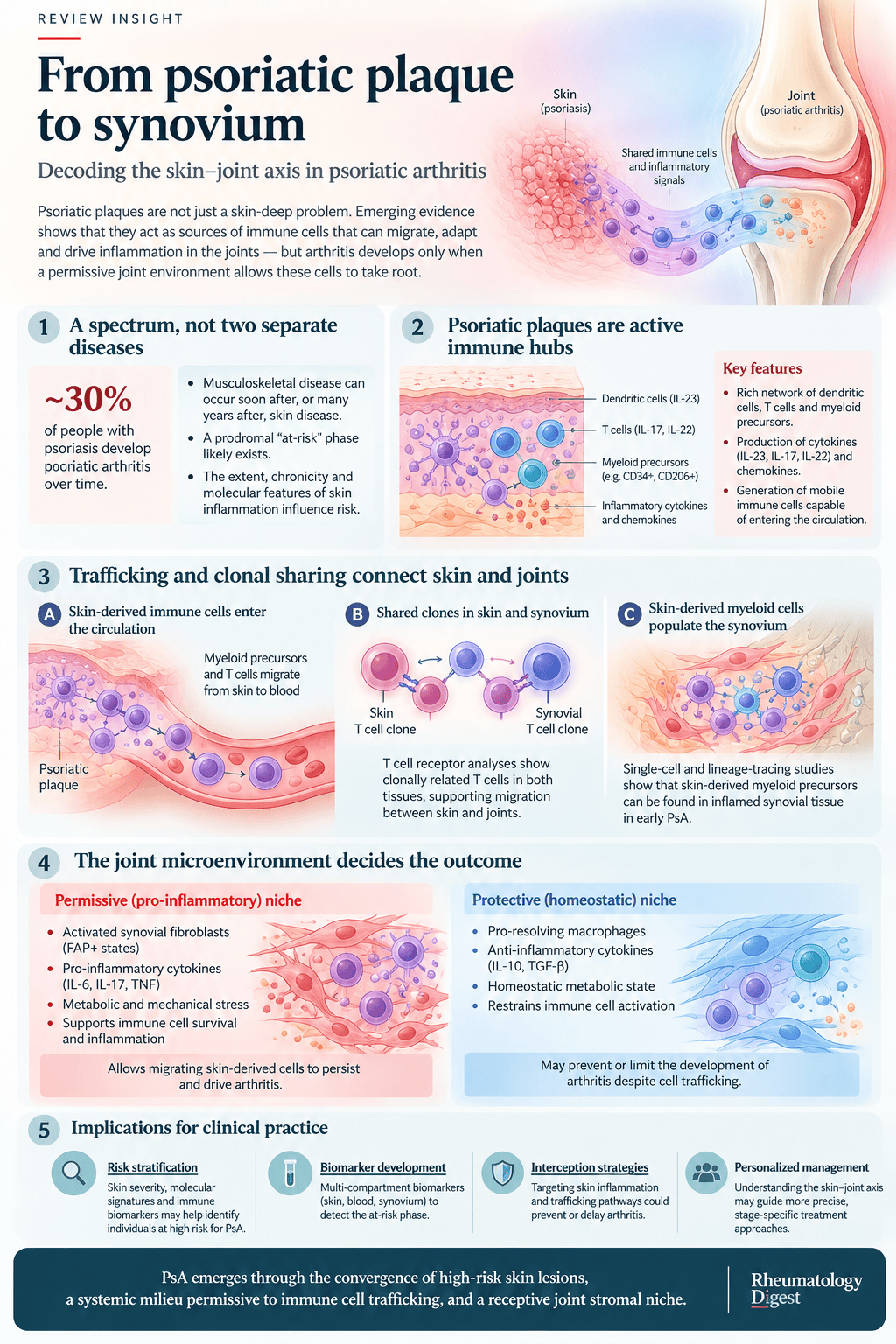
TL;DR: Psoriatic plaques export pathogenic T cells and myeloid precursors to the joint, but arrival is not enough — whether arthritis develops depends on the stromal niche the cells land in, with synovial fibroblasts acting as the gatekeepers.
Psoriatic arthritis develops in up to 30% of people with psoriasis, and the clinical question that follows is the one this review tries to answer mechanistically: why that subset, and why then? The authors’ answer is a three-part model. Plaques generate and export immune cells; the circulation has to be permissive to their travel; and the joint or enthesis has to be receptive when they arrive. The third part is where most of the new evidence sits, and it reframes psoriatic arthritis as a tissue-to-tissue transition rather than a simple extension of skin disease.
Psoriatic disease as a continuum, not two diseases
Psoriasis and psoriatic arthritis were historically treated as separate entities managed by different specialties and linked mainly by epidemiology. The review argues the evidence now favours a spectrum of psoriatic disease affecting skin, peripheral joints, entheses, the axial skeleton and structures outside the musculoskeletal system. Three observations support this. Genetic risk architecture is shared. The cytokine pathways overlap, above all the IL-23–IL-17 axis and TNF. And the same drugs work across domains: TNF inhibitors, IL-17 inhibitors and IL-23 pathway inhibitors are all effective for both skin and joint disease.
Treatment response also shows why the two should not be treated as biologically identical. The skin response often exceeds the musculoskeletal response, and efficacy is discordant between tissues for some targeted therapies. The review treats this as evidence that shared inflammatory pathways are shaped by tissue-specific stromal, immune and biomechanical factors. In this spectrum model the skin is usually, but not invariably, the first clinically visible domain, with joints and entheses activated in sequence or in parallel, which would help explain the clinical heterogeneity.
The clinical picture the biology has to explain
Cumulative incidence of PsA in longitudinal psoriasis cohorts commonly converges on up to 30%, though estimates vary with study design, population and the criteria used. The timing is highly variable: joint disease can appear around the same time as skin disease or years to decades later. That variability supports a prodromal or at-risk phase in which people with psoriasis report intermittent arthralgia, morning stiffness, fatigue or focal entheseal pain without meeting PsA criteria.
Imaging adds weight to that idea. Subclinical inflammation at entheses and periarticular structures seems to precede and predispose to PsA. In a prospective cohort of people with psoriasis, 21.1% developed PsA over a mean follow-up of 28.2 months, and those with baseline structural entheseal lesions carried a markedly higher risk: an incidence of 21.4 versus 4.2 per 100 patient-years, with an adjusted hazard ratio of 5.10.
Clinical features associated with progression include nail disease, scalp involvement, intergluteal or perianal psoriasis, and severe or extensive plaque burden, as well as subclinical enthesitis. But these features do not reliably predict who will progress. The authors suggest they may instead mark tissue patterns more tightly associated with joint pathology. Obesity and metabolic syndrome also raise risk, pointing to metabolic stress, adipose-driven inflammation and mechanical loading as modifiers that lower the threshold for entheseal and synovial inflammation. The practical implications they draw are proactive musculoskeletal screening in higher-risk psoriasis phenotypes, and tighter dermatology–rheumatology integration to cut diagnostic delay, which is associated with worse outcomes.
Why prediction is still poor
Current risk scores for the transition rely largely on clinical features such as pain, imaging findings such as synovitis or enthesitis, and inflammatory markers such as CRP. The review’s criticism is that these may capture a point in time when PsA has already developed, so true mechanistic biomarkers of the transition are missing.
A robust framework, they argue, needs three interlocking axes: the skin-to-blood trafficking of pathogenic cells; the systemic milieu and how permissive it is to trafficking and tissue entry; and joint stromal “gatekeeping”, which decides whether arriving cells become transient passengers, short-lived responders or self-sustaining drivers of chronic inflammation.
Three frameworks for linking skin and joint
The review sets out three non-exclusive ways to conceptualise the connection, and is candid that none is complete alone.
Systemic soluble mediators. Psoriatic plaques, in this view, act as a chronic source of circulating mediators that condition joints from a distance. The IL-23-driven cytokines of psoriasis — IL-17A, IL-17F, TNF and IL-22 — sustain a systemic inflammatory milieu. That milieu could affect joint-relevant microenvironments in several ways: endothelial activation, with increased adhesion molecules such as ICAM1 and VCAM1 and altered vascular permeability that facilitates leukocyte trafficking; priming and reprogramming of resident stromal cells in synovium and entheses, creating a pro-inflammatory “set point”; and amplification of responses to biomechanical stress at the enthesis. Cardiometabolic comorbidity feeds this loop, since adipose-derived mediators, dyslipidaemia, insulin resistance and vascular dysfunction all intensify systemic inflammation, and neuroendocrine inputs such as stress-axis activation and autonomic imbalance may add to it. The limitation they stress: soluble mediators alone cannot explain why only some people with a similar systemic burden develop joint disease. Tissue-specific gatekeeping must still determine whether arthritis starts and persists.
Shared predisposition. Here psoriasis and PsA are two expressions of one underlying condition, with a common genetic background triggered by environmental factors such as mechanical stress, infection and obesity. Genome-wide studies have found loci in both HLA and non-HLA genes, including polymorphisms in IL-23–IL-17 pathway genes and in NF-κB regulators such as TNFAIP3. Some loci seem to define susceptibility to particular phenotypes, such as nail disease or a predominantly axial or peripheral pattern. Epigenetics may contribute: a genome-wide methylation study in people with early psoriasis identified markers that distinguished those who later developed PsA, with good accuracy, suggesting the systemic immune programme could be established before musculoskeletal symptoms appear. In 2026, epigenetic remodelling was also linked to rheumatoid arthritis progression in anti-CCP-positive individuals. The authors regard this framework as largely descriptive.
Interactions with other barrier tissues. Psoriatic disease may be a disorder of interconnected barrier tissues rather than skin and joints in isolation. The gut–joint axis is established in spondyloarthritis, and a skin–gut–joint axis is plausible in psoriasis. A mouse model with a Card14 variant shows gut microorganisms driving skin inflammation through a metabolic relay in which indole-producing bacteria promote host indoxyl sulfate production and amplify cutaneous Th17 responses, and serum indoxyl sulfate correlates with disease severity in people with psoriasis. The skin remains uniquely accessible for serial sampling, which makes the psoriasis-to-PsA transition a natural human model for tissue-to-tissue spread. The relative contributions of the gut and skin axes to progression remain unclear.
T cells that move between skin and joint
The most concrete evidence for a physical link comes from T cell receptor sequencing.
Clonal sharing. Early studies using TCR profiling and CDR3 sequencing found overlapping oligoclonal expansions between lesional skin and synovium in individual PsA patients, including identical or highly similar CDR3 sequences, which suggested antigen-driven responses spanning both tissues. Single-cell RNA sequencing with paired αβ TCR analysis has since provided quantitative confirmation: sizeable fractions of the synovial CD8+ repertoire can be traced to clones shared between skin and joint, which often keep a tissue-resident, cytotoxic transcriptional programme and sit in antigen-presenting-cell neighbourhoods in both tissues. Complementary datasets show an intermediate step in blood. Circulating CCR4+CD8+ central memory cells, with a skin-homing phenotype, share clonotypes with differentiated CD8+ effector cells in synovial fluid, which offers a plausible systemic link between priming in the plaque and effector accumulation in the joint. The shared clones usually show a type 17 profile, expressing IL-17A, IL-17F and GM-CSF, and often tissue-residency features such as CD69; one subset expresses granzyme K, associated with complement-driven amplification of inflammation. Entheseal data are still missing.
Tissue-resident memory cells that do not stay put. Tissue-resident memory (T_RM) cells were defined as long-lived, non-recirculating sentinels anchored in barrier tissues by sustained residency programmes. In psoriasis they are implicated in persistence and in site-specific relapse, acting like an “immunological scar” that rapidly drives inflammation when a trigger recurs. The new finding is that T_RM states are not fixed. Under heightened inflammation, some of these cells can lose residency constraints, re-express egress-associated markers, enter the bloodstream as “ex-T_RM” cells, and re-home to inflamed or permissive niches. If so, plaque T_RM cells would be more than local amplifiers; they would be a reservoir of primed T cells able to seed distant sites including the joint. The authors say clear evidence for circulating T_RM cells in psoriatic disease is still lacking.
Circulating CD8+ effectors in a humanised mouse model. In NSG-SGM3 mice reconstituted with blood cells and serum from PsA patients, circulating human CD8+ effector cells accumulated in skin and synovium and reproduced both psoriasiform skin changes and joint features including dactylitis, enthesitis and erosive disease. Depleting CD8+ T cells attenuated both skin and synovial disease, and immunoglobulin-containing serum was required for full expression, suggesting humoral factors cooperate with trafficking cells. The authors are careful that this does not prove synovial effector cells originate in plaques, and that the cohorts involved are small and need validation.
What this means for the transition. One possibility the review raises is that most people with psoriasis have skin-primed T_RM or cytotoxic T cells that periodically circulate and survey peripheral tissues, but never meet a permissive environment or antigen. In those who develop PsA, arrival in a niche primed by mechanical stress, stromal activation, or microbial antigen presentation produces a permissive inflammatory milieu, and the T cells then cooperate with myeloid cells and fibroblasts to sustain chronic synovitis. They also note that the co-occurrence of clonal CD8+ populations in plaque and joint is not formal proof of migration, and the same cells could in theory arise from gut priming or secondary lymphoid organs.
Myeloid cells: the direct trafficking evidence, from mice
The strongest direct demonstration of skin-to-joint movement comes from IL-23-driven mouse models, which are the experimental basis for the central role of IL-23. Skin-specific or systemic IL-23 overexpression, or repeated cutaneous injection, recapitulates psoriasis with an IL-17-dominated inflammation and, in some settings, produces enthesitis and arthritis, with enthesitis an early musculoskeletal manifestation. These models have supported the clinical success of IL-23 and IL-17 inhibitors, and IL-17-producing cells have been identified in the affected joints of IL-23-overexpressing mice. The model comes with a caveat: the dichotomous skin-versus-joint phenotype depends on strain, with the C57BL/6 background developing skin disease but appearing resistant to joint involvement. The authors see that as an opportunity, since it mirrors the heterogeneity in humans.
Lineage tracing. In IL-23-overexpressing reporter mice, photoconversion allowed cells in inflamed skin to be permanently labelled and tracked. Labelled cells later appeared in articular and entheseal compartments. Single-cell analysis identified a CD2+MHCII+ myeloid precursor population as the key migratory cell. Crucially, arrival of these cells in synovial or entheseal tissue was not sufficient to cause arthritis. They also reached the musculoskeletal tissue of arthritis-resistant mice, which indicates that skin-to-joint migration may be a broad feature of psoriatic disease rather than a predictor of joint inflammation. What differed was the recipient niche. In arthritis-prone mice the skin-derived cells became pro-inflammatory macrophages in synovium or enthesis; in resistant mice the same precursors were skewed toward an anti-inflammatory phenotype associated with protection and resolution.
Human correspondence. Single-cell RNA sequencing, imaging mass cytometry and mitochondrial DNA lineage tracing in paired tissue from people with psoriasis and early PsA identified myeloid precursors expressing CD2, MHCII and CCR2 in psoriatic skin, with transcriptional features similar to those in the IL-23 mouse model. Spatial analysis located them in synovial niches close to fibroblasts and stromal elements and T cells, with different patterns in non-arthritic tissue. Mitochondrial lineage tracing showed that myeloid lineages, particularly the CD2+MHCII+CCR2+ precursors, carried shared somatic variants across skin and synovium, direct evidence that a subset of synovial myeloid cells is clonally related to cells in psoriatic skin. The conclusion the authors draw: there is a functional myeloid arm of the skin–joint axis, but local tissue permissiveness, not migration alone, probably determines progression to clinical arthritis.
Fibroblasts as gatekeepers
This is the review’s central argument. Synovial and entheseal fibroblasts can be thought of as the gatekeepers of the axis, deciding whether skin-derived immune cells are converted into arthritis or redirected towards protection and repair.
Heterogeneity. Single-cell and spatial studies in RA and PsA have identified distinct fibroblast subsets, including lining-layer fibroblasts associated with cartilage and bone-adjacent tissue remodelling, and sublining populations that orchestrate immune recruitment and vascular remodelling. Pathogenic subsets often express fibroblast activation protein (FAP) and podoplanin together with pro-inflammatory mediators, and are enriched at pannus–bone interfaces and perivascular niches. Fibroblast populations rich in regulatory molecules such as CD200 and other anti-inflammatory mediators seem to support tissue homeostasis and resolution. RA and PsA appear to share broad fibroblast archetypes, but differences in subset abundance, activation and distribution might underlie disease-specific patterns of joint damage and extra-articular involvement. Understanding of fibroblast subsets in PsA is considerably thinner than in RA.
Fibroblasts shape myeloid fate. Through direct contact, extracellular matrix components and secreted factors, fibroblasts direct monocytes and myeloid precursors towards macrophage states ranging from highly pro-inflammatory and tissue-destructive to pro-resolving and reparative. In the models described, abundant CD200+ fibroblasts push incoming myeloid cells toward anti-inflammatory, tissue-protective profiles, whereas expansion of FAP+ pathogenic fibroblasts favours pro-inflammatory macrophage differentiation, which sustains synovitis and promotes structural damage.
Permissive versus protective niches. From this follows the distinction at the heart of the review. A permissive niche, with FAP+ fibroblasts, pro-inflammatory macrophages, altered metabolic state and inflammatory cytokines, retains, reprograms and amplifies incoming immune cells, leading to synovitis and enthesitis. A protective niche, with CD200+ fibroblasts and other pro-resolving fibroblasts, balanced matrix and regulatory cytokine profiles, diverts skin-derived myeloid cells to reparative roles or apoptosis, and may skew responding T cells toward regulatory phenotypes. The authors suggest such niches vary between individuals and between joints, offering a plausible explanation for the incomplete and asymmetric progression from psoriasis to PsA.
What it adds clinically. It reinterprets heterogeneity. A joint with a largely protective stromal composition might resist arthritis even with robust skin inflammation and demonstrable trafficking. Conversely, in someone with a permissive stromal profile — shaped by genetics, age, expanded pro-inflammatory CD8+ clones, mechanical loading or previous inflammation — modest trafficking might be enough to establish persistent synovitis. That fits previous findings that joint-specific memory resides in synovial macrophages, synovial fibroblasts and T_RM cells.
What the skin tells you about joint risk
Only a minority of people with psoriasis develop PsA, which, the review argues, speaks against a model based purely on systemic inflammation and favours a multistep process in which skin, circulation and the joint niche must align.
At the skin level, plaques are biologically heterogeneous. Differences in T_RM and Tc17 cell activity, myeloid organisation, cytokine output, vascular remodelling, local microanatomy and microbiota probably determine how efficiently a lesion produces “exportable” pathogenic cells. That might explain why nail disease, scalp involvement and intergluteal or perianal psoriasis correlate more strongly with PsA risk, consistent with a distinct biomechanical and immunological milieu. Metabolic stress may further amplify it. The bloodstream then works as a conduit, and circulating cytokines and chemokines can promote endothelial activation with adhesion molecules and chemokine presentation that support tissue entry.
The authors’ composite statement: PsA emerges when three features converge — skin-derived immune cells with musculoskeletal homing potential, a systemic milieu permissive for trafficking, and a receptive synovial or entheseal niche that stabilises chronic inflammation. They are explicit that the framework needs longitudinal, multisite validation.
When the joint comes first
An important caveat to a skin-first model: in an estimated 10–15% of patients, musculoskeletal inflammation precedes or arises at the same time as psoriasis. The review does not treat these as exceptions that break the model, and offers several non-exclusive readings. They may still follow a skin–joint trajectory with the skin component clinically occult — subclinical inflammation, microscopic barrier dysfunction, or hidden sites such as scalp, nails and intergluteal or perianal skin that are insufficient for diagnosis but able to prime pathogenic cells. The joint might simply be the first tissue to cross a clinical threshold, perhaps because entheseal microdamage and biomechanical stress create a permissive niche that amplifies otherwise modest signals. Or shared upstream triggers, genetic risk, and microbial or barrier-derived signals could activate parallel “tissue modules” in skin and joint, with stochastic dominance of one compartment. A further possibility is a stronger contribution from circulating T_RM cells that are skin-imprinted but no longer skin-resident, seeding joints before skin disease is visible; in some patients PsA might even arise from a predominantly joint-driven or extra-cutaneously primed pathway. The practical message is that paired, multicompartment profiling — including careful assessment of nails and hidden skin sites — is needed to tell these apart.
Biomarkers, imaging and intercepting the transition
The proposed translation is multi-compartment. In skin, transcriptomic or proteomic signatures of activated T_RM and Tc17 cells and enrichment of skin-derived myeloid precursors could be obtained from lesional biopsies or minimally invasive methods such as tape stripping, to identify “PsA-prone” signatures within plaques. In blood, quantitative and phenotypic profiling of skin-primed T cells and myeloid precursors, with relevant cytokines and chemokines, might mark trafficking and systemic permissiveness. At the synovium or enthesis, advanced imaging such as FAP tracers and, where appropriate, biopsy-derived samples could characterise stromal gatekeeping.
For clinical practice the review proposes: structured musculoskeletal screening and targeted imaging or biomarker assessment in dermatology clinics for high-risk phenotypes, to detect imminent PsA earlier; rheumatology evaluation of people with psoriasis and nonspecific arthralgia that integrates biomarker profiles and imaging indicators of an active skin–joint axis; and dedicated joint dermatology–rheumatology “at-risk PsA” clinics as a platform for prevention approaches.
On intervention, the multilayer model points to rational targets: controlling cutaneous inflammation, particularly through IL-23–IL-17 pathway modulation; reducing systemic permissive cues, including metabolic risk; and, looking ahead, reshaping synovial stromal niches towards protective, pro-resolving states — for example by enhancing CD200+ fibroblast networks or constraining pathogenic FAP+ subsets. Next-generation trials will need to pair clinical endpoints with mechanistic readouts across skin, blood and joint, to show control and genuine modification of the transition from psoriasis to PsA.
The future-directions section calls for blood- or serum-based markers that reflect either joint-related or skin-related risk, minimal biomarker panels integrated with imaging features (subclinical synovitis, enthesitis or tenosynovitis on ultrasound or MRI), able to identify high-risk individuals over a clinically actionable window such as 1–3 years. Those panels must also account for the temporal stability of peripheral immune signatures and how conventional or targeted therapy alters them. Simplified lineage-tracing experiments translated into longitudinal human studies could provide indirect evidence of skin-to-joint trafficking without prohibitive cost, and well-characterised psoriasis cohorts are the place to start.
What to hold loosely
Several features of the evidence deserve caution, most flagged by the authors themselves.
- The direct trafficking evidence for myeloid cells comes from mice. The human data show clonal relationships and spatial colocalisation across tissues, which is strong, but they cannot show direction or timing of movement.
- The T cell findings come from relatively small cohorts, and the authors say they need validation in larger independent datasets.
- The circulating T_RM concept is plausible but unproven, and the clonal co-occurrence is not formal proof of migration.
- The stromal gatekeeper model is largely extrapolated from RA fibroblast biology and the mouse data, with PsA-specific fibroblast subsets much less defined.
- A skin–joint axis does not mean biological equivalence of psoriasis and PsA. The variable latency between skin and joint disease, the incomplete transition, and the discordant efficacy across tissues all point to real divergence.
- The overall framework needs longitudinal, multisite validation, and the translational proposals — at-risk clinics, biomarker panels, stromal niche reprogramming — are proposals, not tested interventions.
What it adds up to
The review’s contribution is a change of question. The older one asked how psoriasis becomes PsA as though it were a single process of spread. The newer one asks why immune cells that reach many joints cause arthritis in only some. The proposed answer — that the receiving tissue, and specifically its fibroblast and macrophage composition, decides — has two practical consequences the authors draw out. For prediction, the useful biomarkers will likely need to capture skin, blood and joint together rather than any one compartment. And for prevention, the most interesting long-term idea is not suppressing the skin or the trafficking cells harder, but reprogramming the joint’s stromal niche toward a protective state — an approach that remains, for now, a hypothesis rather than a therapy.
Disclosures noted in the source: the authors declare no competing interests. Several authors are supported by German Research Foundation, European Research Council and other public grants listed in the paper.