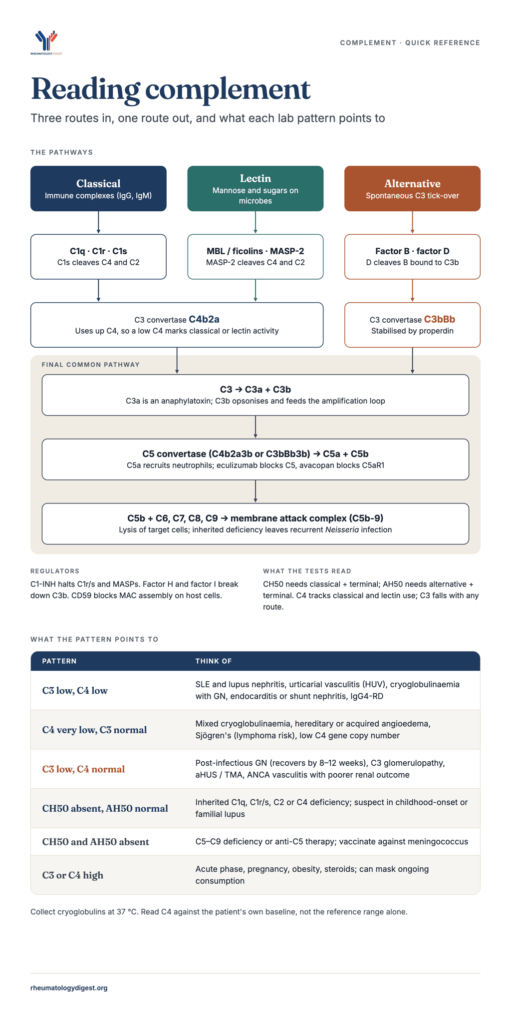
TL;DR: Read complement by pattern, not by number: low C3 with low C4 points to immune-complex consumption, low C3 with a normal C4 to the alternative pathway, and a very low C4 with a normal C3 to cryoglobulins, C1-inhibitor failure or a constitutionally low C4 gene copy number, and the functional assays (CH50 and AH50) locate inherited or drug-induced defects.
Quick reference: start from the pattern
Find the row that matches the result, then follow the link for prognosis and work-up. The infographic above sets the pathways out in one picture.
| Pattern | Pathway implied | Main causes | Useful next tests |
|---|---|---|---|
| Low C3 and low C4 | Classical (immune complex) consumption | SLE / lupus nephritis, HUV, mixed cryoglobulinaemia with active GN, serum sickness, endocarditis and shunt nephritis, IgG4-RD | dsDNA, anti-C1q, cryoglobulins (at 37 °C), blood cultures, IgG4, urine and biopsy |
| Low C4, normal or mildly low C3 | Early classical (C1 step) | Mixed cryoglobulinaemia, HAE / acquired angioedema, Sjögren’s (lymphoma risk), low C4 copy number, quiescent SLE | Cryoglobulins, RF, HCV/HBV, C1-INH level and function, C1q, serum free light chains |
| Low C3, normal C4 | Alternative pathway / amplification loop | Post-infectious GN, C3 glomerulopathy, aHUS / TMA, partial lipodystrophy, AAV with poor prognosis, C3 or factor H/I deficiency | Repeat C3 at 8–12 weeks, C3NeF, factor H/I, anti-factor H antibody, genetics, monoclonal screen if over 50 |
| CH50 absent, AH50 normal | Early classical deficiency | C1q, C1r/s, C2, C4 deficiency (SLE risk) | Individual components |
| CH50 and AH50 both absent, C3 and C4 normal | Terminal deficiency (or anti-C5 drug) | C5–C9 deficiency, eculizumab / ravulizumab | Individual C5–C9; Neisserial vaccination |
| AH50 low, CH50 normal | Alternative pathway factor deficiency | Factor B, D or properdin deficiency | Individual factors |
| Raised C3 / C4 | Acute phase synthesis | Inflammation, infection, pregnancy, obesity, steroids | None; consider whether consumption is being masked |
Where to read more for each pattern
- Low C3 and low C4: SLE and lupus nephritis, cryoglobulinaemia and urticarial vasculitis, and IgG4-related disease and APS.
- Low C4 with normal or mildly low C3: isolated low C4, cryoglobulinaemia and Sjögren’s disease.
- Low C3 with normal C4: isolated low C3 and ANCA-associated vasculitis.
- Abnormal CH50 or AH50: inherited deficiencies and infection risk.
- Raised C3 or C4, odd or discordant results, or a patient on a complement inhibitor: raised complement, laboratory pitfalls and therapy.
Test cheat sheet
| Test | What it measures | How to read it |
|---|---|---|
| C3 and C4 | Protein concentration, usually by nephelometry or turbidimetry | The balance between consumption and synthesis. Both are acute phase reactants, so inflammation can lift a consumed level back into the reference range. Read C4 against the patient’s own baseline |
| CH50 | The whole classical pathway, C1 to C9 | Absent with a normal AH50 points to an early classical component (C1, C2, C4) |
| AH50 | The alternative pathway together with C3 and C5–C9 | Absent with an absent CH50 points to C3 or a terminal component, to global consumption, or to anti-C5 therapy |
| Anti-C1q | Follows renal rather than global activity in lupus | Sensitivity 74% and specificity 77% for active nephritis, and a useful negative predictive value. Also classic in hypocomplementaemic urticarial vasculitis |
| C1q, C1-inhibitor level and function | Separates the C1-inhibitor and C1q disorders | Low C1q with a normal C1-inhibitor suggests HUV. Low C1q with low C1-inhibitor suggests acquired angioedema. Normal C1q with low C1-inhibitor suggests HAE |
| C3NeF, factor H and I, anti-factor H antibody | Alternative pathway dysregulation | C3 can be normal even when C3NeF is present, so a normal C3 should not stop the work-up |
| Cell-bound C4d and split products (for example iC3b/C3) | Complement activation | Outperform plain C3 and C4 for lupus activity, but access is limited outside the US |
| Cryoglobulins | Cold-precipitating immunoglobulin | Collect at 37 °C. A single negative test does not exclude cryoglobulinaemia when the complement pattern fits |
Before you interpret a result
- Check how the sample was handled. Cryoglobulins need a pre-warmed plain tube kept at 37 °C. Serum left at room temperature can lower CH50 and C4 artefactually, and functional assays and split products need prompt separation and freezing at −70 °C.
- Check the timing. Draw samples before plasma exchange, IVIG or the first dose of a complement inhibitor, because afterwards CH50 and AH50 reflect the drug.
- Look for low levels without consumption. Liver failure, malnutrition, nephrotic syndrome, protein-losing enteropathy, plasma exchange and a low C4 gene copy number all lower complement without immune activity.
- Remember what raises it. Acute phase response, pregnancy, obesity, oestrogen and steroids lift C3 and C4, and active inflammation can mask consumption.
- Use the patient’s own baseline. A permanently low C4 with no sign of consumption may simply reflect gene dosage.
The sections below give the prognostic and work-up detail behind each row of the table.
How a complement result is read
The pathway behind an abnormal result matters more than the fact that complement is low. Which components are depleted, and whether a functional assay is affected, separates immune-complex consumption, alternative-pathway dysregulation, C1-inhibitor failure and inherited deficiency. Each points to a different differential and a different next test.
C3 and C4 are measured as protein concentrations, usually by nephelometry or turbidimetry. CH50 tests the whole classical pathway from C1 to C9, and AH50 tests the alternative pathway together with C3 and C5–C9 (Mayo Clinic Laboratories). Interpreting the two functional assays together locates a defect. A low CH50 with a normal AH50 points to an early classical component (C1, C2, C4). Both being absent points to C3 or a terminal component, or to global consumption (Mayo AH50 interpretation).
A low level reflects the balance between consumption and synthesis. Complement can therefore fall without any immune activity: in liver failure, malnutrition and protein-losing states, through genetic copy-number variation, or from in vitro activation after collection. C3 and C4 are also acute phase reactants. Inflammation can lift a consumed level back into the reference range, which is part of why their sensitivity for flares in SLE is only modest.
Two rules of thumb follow. A persistently low C4 with a normal C3 is mostly classical-pathway activation at the C1 step (cryoglobulins, C1-inhibitor deficiency) or a low C4 gene copy number. A low C3 with a normal C4 means the alternative pathway or the amplification loop until proven otherwise.
SLE and lupus nephritis
In SLE, low C3 and C4 support classification and signal activity, but they are imperfect monitors of flares. The 2019 EULAR/ACR criteria give 3 points for low C3 or low C4 and 4 points for both together (Aringer 2019). A 2025 cohort supported the higher weight for both being low: those patients had more cutaneous, haematological and serological activity (Front Med 2025). Hypocomplementaemia is present at diagnosis in about 50–89% of patients, so a normal level never excludes lupus (Ayano & Horiuchi).
Flare prediction is modest. The Ohio SLE Study measured C3 and C4 every two months in 71 patients with lupus nephritis, who had 70 renal flares. Sensitivity for flare was 75% for C3 and 48% for C4, and neither level reliably fell before the flare (Birmingham 2010). A systematic review informing treat-to-target examined patients who were clinically quiescent but serologically active. Low C3 during stable disease carried a 2.0–3.8-fold flare risk in three of seven studies, and low C4 predicted flares in three of four (Lupus 2022 SLR). The practical reading is closer follow-up for serologically active, clinically quiet patients, not automatic escalation.
Prognosis in nephritis. In a 445-patient Italian multicentre cohort, isolated low C3 (≤80 mg/dL with normal C4) six months after biopsy roughly doubled the risk of CKD, ESKD or death. A rising C3 over those six months was protective (Andrulli 2025). Persistent low C3 despite treatment suggests ongoing alternative-pathway activity or a concurrent TMA, and should prompt a review of the biopsy.
Anti-C1q antibodies follow renal rather than global activity. In a meta-analysis, sensitivity for active versus inactive nephritis was 74% and specificity 77% (Yin 2012, via Quest). The negative predictive value is the most useful property. In one cohort, none of the patients who were anti-C1q negative developed class IV or V nephritis (NPV 100%, 18/18) (Meyer 2009).
Activation products outperform levels. Erythrocyte- and B-cell-bound C4d (CB-CAPs) were abnormal in 62% of patients with SLE, against 38% with low C3/C4 (Lupus Sci Med 2020). In paediatric SLE, B-cell C4d gave an AUC of 0.91, compared with 0.63 for C3 and 0.67 for C4 (paediatric CB-CAPs cohort). Plasma ratios of split product to parent, such as iC3b/C3, also separate flaring from non-flaring disease better than C3 or C4 (Curr Rheumatol Rep 2021). Access to these tests is limited outside the US.
Inherited deficiency of an early classical component is the strongest single genetic risk for lupus. About 90% of people with complete C1q deficiency develop SLE, as do 65–80% of those lacking C1r, C1s or C4. Complete C2 deficiency (around 1 in 20,000) carries roughly a 25% penetrance (Arthritis Rheumatol 2022). Suspect it when lupus starts in childhood, involves both sexes equally, is dominated by skin disease, comes with recurrent encapsulated-organism infection, or shows an undetectable CH50 with near-normal C3. Low C4 gene copy number (C4A especially) is common and can keep C4 permanently low without any activity. That is one reason to judge a patient’s C4 against their own baseline.
Cryoglobulinaemia, urticarial vasculitis and other immune-complex vasculitides
Mixed cryoglobulinaemia (types II and III) gives the most disproportionate pattern in rheumatology. C4 is very low or undetectable, CH50 is low, and C3 is normal or only mildly reduced (Rev Esp Enferm Dig). Monoclonal or polyclonal IgM rheumatoid factor bound to IgG fixes C1q and uses up C4 and C2 early in the cascade. C4 often stays low in clinical remission, so it tracks activity poorly. A clearly low C3 usually accompanies active membranoproliferative GN.
Low C4 still carries prognostic weight. In the EuroCryo cohort of 1,294 patients, it independently predicted severe events (hazard up 38%) together with RF positivity, older age, male sex and higher creatinine (Saadoun 2026, Rheumatology; EMJ summary). Low C4 was also linked to lymphoma, as was autoimmune aetiology, with Sjögren’s the commonest cause at 21%. In the direct-acting antiviral era, HCV aetiology was protective (HR 0.74).
Hypocomplementaemic urticarial vasculitis (HUV, McDuffie syndrome) combines urticarial lesions lasting more than 24 hours, leukocytoclastic vasculitis and low complement. It is classically associated with anti-C1q antibodies (Marzano 2022, JACI). In a French national series of 57 patients, the usual finding was low C1q with normal C1-inhibitor; anti-C1q was positive in 55%. Angioedema occurred in 51%, with frequent musculoskeletal and ocular involvement (Jachiet 2015). Renal disease is less common but can be MPGN or crescentic, and carries a worse prognosis (Ion 2020, JCM). Three practical points follow. First, normocomplementaemic urticarial vasculitis is usually skin-limited. Second, HUV overlaps with SLE, so test dsDNA and Sm. Third, a low C1q with a normal C1-inhibitor separates HUV from acquired angioedema, where both are low.
Serum sickness and drug- or infection-triggered immune-complex disease (classically heterologous antisera or anti-thymocyte globulin, and serum-sickness-like reactions to antibiotics) can lower C3 and C4 transiently during the acute phase. Recovery within weeks, once the antigen is cleared, supports the diagnosis.
Rheumatoid vasculitis and Felty syndrome are the settings where complement falls in RA. In uncomplicated RA, C3 and C4 are typically normal or raised as acute phase reactants. New low complement in a patient with long-standing seropositive RA should prompt a search for vasculitis or cryoglobulins.
Other rheumatic diseases where complement carries prognostic weight
Sjögren’s disease. Low C4 is the most reproducible serological predictor of lymphoma and death. In Ioannidis’ Greek cohort of 723 patients, low C4 at the first visit predicted lymphoproliferative disease and mortality (HR 4.39 for death). Low C4 together with palpable purpura defined a high-risk “type I” phenotype, present in about 21% of patients (Ioannidis 2002). In Theander’s population-based cohort, low C3 (HR 6.2) and low C4 (HR 9.5) predicted lymphoma, and the standardised incidence of NHL was about 16-fold (Theander 2006, ARD). Low complement at diagnosis should move a patient onto closer surveillance: glands, nodes, purpura, cryoglobulins, monoclonal band and β2-microglobulin.
IgG4-related disease. Hypocomplementaemia occurs in 20–40% of patients overall and in 50–70% of those with kidney involvement (IgG4-RKD biopsy study). It travels with high IgG, high IgG4 and high non-IgG4 IgG, and with nodal, lung and renal disease. In a Japanese cohort it independently predicted relapse (OR 6.8) (Fujita 2022). A European kidney cohort found the same link with relapse, and also found that first-line rituximab was protective (European IgG4-RKD cohort, CJASN 2023). Unexplained hypocomplementaemia with tubulointerstitial nephritis and raised IgG should bring IgG4-RD into the differential alongside lupus nephritis and Sjögren’s TIN.
ANCA-associated vasculitis. Overt hypocomplementaemia is unusual, but C3 at diagnosis carries prognostic information. In the Parma cohort, C3 was below the lower limit in 35% of patients and C4 was low in only 2%. The low-C3 group had worse death-censored renal survival and more often had histological TMA (Manenti 2015, CJASN). A C3 in the lower half of the normal range already predicted worse outcomes in other cohorts (Augusto 2016; Crnogorac 2018). These findings are consistent with alternative-pathway and C5a involvement, which is the rationale for avacopan.
Antiphospholipid syndrome. Low C3 and C4 occur in a minority and point to complement-mediated injury. In the EUROAPS registry of 1,048 women with obstetric APS, 21% had low complement during pregnancy. Those women delivered earlier, had fewer live births, and had more fetal loss when double or triple aPL-positive (Esteve-Valverde 2023, Placenta). Earlier studies disagree on this (Front Immunol 2020 table), so low complement is a risk-stratifying flag rather than a treatment trigger. In catastrophic APS or APS nephropathy with TMA, falling C3 supports complement-driven microangiopathy (see the TMA paragraph under isolated low C3).
Isolated low C3: alternative-pathway disorders and infection-related GN
A low C3 with a normal C4 in a patient with nephritis is the alternative pathway at work until proven otherwise. The main question is whether it is a self-limiting infection-related process or ongoing dysregulation.
Post-infectious (post-streptococcal) GN. The usual pattern is low C3 with normal C4, and C3 recovers within 6–8 weeks in most patients (BMC Nephrol 2024). A low C3 at 6–8 weeks warrants reassessment. Persistence beyond about 12 weeks is a red flag for C3 glomerulopathy unmasked by infection (Rev Nefrol 2026). Some genuine post-streptococcal GN still recovers late, so if the patient is clearly improving, a biopsy can be deferred (Buffalo series).
C3 glomerulopathy (dense deposit disease and C3GN). Most patients have persistently low C3 with normal C4 (Front Med 2023). The cause is mostly autoimmune, through C3, C4 or C5 nephritic factors (about 65%), and less often genetic (about 20%) (Iowa MORL). C3 can be normal even when C3NeF is present, so a normal C3 should not stop the work-up (J Clin Pathol 2024). For patients over about 50, screen for a monoclonal gammopathy (“monoclonal gammopathy of renal significance”). Iptacopan, an oral factor B inhibitor, was approved by the FDA for C3G in March 2025. Pegcetacoplan, a C3 inhibitor, followed in July 2025 for C3G and primary IC-MPGN in patients aged 12 and over (HCPLive).
Partial lipodystrophy. Acquired loss of fat from the face and upper body, alongside C3NeF and low C3, often precedes dense deposit disease. C3NeF-driven lysis of adipocytes that express factor D is thought to be the mechanism (Front Med 2023). The pattern can appear in patients with SLE or dermatomyositis.
Atypical HUS and complement-mediated TMA. C3 is low in only a subset, so a normal C3 does not exclude aHUS. Reported frequencies are 30–50% with CFH variants, 70–80% with C3 variants and 40–60% with anti-factor H antibodies (OJRD table). In India, anti-factor H antibody disease accounts for about half of paediatric aHUS, mainly in children aged 5–15. The Indian Society of Pediatric Nephrology recommends sending anti-FH antibody, C3 and CD46 samples before plasma exchange (Indian Pediatr 2020). For rheumatologists, the overlap matters most in SLE, APS, scleroderma renal crisis and AAV with histological TMA. In AAV, TMA tracked with low C3 and nearly universal ESKD (Manenti 2015).
Infective endocarditis and shunt nephritis. These are important mimics of vasculitis. In the largest biopsy series of endocarditis-associated GN, the commonest lesion was necrotising crescentic GN (53%), and staphylococcal tricuspid infection predominated (Boils 2015, Kidney Int). Hypocomplementaemia was found in 56% of those tested and ANCA in 28% (same series). A patient with ANCA, low complement and RPGN needs blood cultures and an echocardiogram before induction immunosuppression. Shunt nephritis follows chronic infection of ventriculoatrial shunts and typically produces MPGN with low complement (KDIGO 2012).
Isolated low C4: C1-inhibitor deficiency and C4 genetics
A low C4 with a normal C3 and no cryoglobulins should raise two possibilities: C1-inhibitor deficiency, and a constitutionally low C4 gene copy number.
Hereditary and acquired angioedema. In C1-inhibitor deficiency, C4 is low in nearly every case, including between attacks. That makes C4 the screening test for recurrent angioedema without urticaria. If C4 and C1-INH antigen are both low on two occasions, the diagnosis is C1-INH deficiency. If only C4 is low, measure C1-INH function (Cicardi & Zanichelli 2010). C1q then separates the two forms. It is normal in HAE and low in about 70% of acquired angioedema, which typically starts after 40 and has no family history. Acquired angioedema is associated with MGUS (48%) and non-Hodgkin lymphoma (27%), so it is effectively a paraneoplastic flag (Allergy Asthma Clin Immunol 2019). It is also reported with SLE and other autoimmune diseases (Int Arch Allergy 2021). For rheumatologists the useful triad is: low C4 and low C1q with normal C1-INH suggests HUV; low C4 and low C1q with low C1-INH suggests acquired angioedema; low C4 with normal C1q and low C1-INH suggests HAE.
C4 copy-number variation. Most people carry two to six C4 genes (C4A and C4B), and a few carry more, so the “normal” serum C4 varies widely. Low total C4 copy number and C4A deficiency are recognised SLE risk factors (Front Immunol 2016). The consequence is that a patient with a permanently low C4 and no sign of consumption (normal C4d, quiet clinically) may simply have low gene dosage. Treating that C4 as a target leads to over-treatment, and serial values against the patient’s own baseline mean more than any single reading.
Inherited deficiencies and infection risk
Which component is deficient predicts which infection. Early classical-pathway defects (C1q, C1r/s, C4, C2) lead to SLE-like disease and infections with encapsulated organisms. C3 deficiency, or C3 depleted by absent factor H or factor I, causes recurrent pyogenic infection and MPGN or aHUS. Terminal (C5–C9) and properdin deficiencies cause Neisserial disease.
Terminal pathway and properdin. Inherited C5–C9 deficiency raises the incidence of meningococcal disease up to 10,000-fold. These infections are often recurrent, with unusual serogroups, and paradoxically less lethal than in people with normal complement (ACP MKSAP). Screen with CH50 (with AH50 for alternative-pathway defects) after recurrent Neisserial infection, any Neisserial infection in more than one family member, or invasive disease with a non-groupable or uncommon serogroup. Confirmed patients need MenACWY conjugate and MenB vaccines.
Iatrogenic deficiency. Eculizumab and ravulizumab (anti-C5), now used by rheumatologists and nephrologists for aHUS, CAPS and refractory TMA, reproduce terminal deficiency. They carry a 1,000–2,000-fold increase in meningococcal risk, and cases still occur after vaccination (CDC MMWR 2017). Vaccinate at least 2 weeks before the first dose, and give antibiotic prophylaxis for the duration of treatment, as UK and French guidance recommends (US eculizumab cohort 2020). Expect CH50 and AH50 to be suppressed on treatment. Iptacopan (factor B) and pegcetacoplan (C3) carry the same encapsulated-organism precautions.
Mannose-binding lectin deficiency is common (genetically low MBL in roughly 5–10% of people) and mostly silent. It is a modifier rather than a disease, linked in some studies to infection during chemotherapy or in infancy. Test with a lectin-pathway assay only when CH50 and AH50 are normal but suspicion of a complement defect remains.
Raised complement, laboratory pitfalls and complement-targeted therapy
Raised C3 and C4 are acute phase responses and rarely have diagnostic meaning. They behave like other positive acute phase proteins in infection and inflammation (Ritchie, J Clin Lab Anal). Obesity, the metabolic syndrome, oestrogen exposure and corticosteroids raise them too. Both climb steadily through normal pregnancy, so a C3 that falls or fails to rise in a pregnant woman with lupus is significant even within the non-pregnant reference range (Perinatology reference). Active inflammation can also mask consumption, keeping a consumed C3 inside the reference range.
Low levels without consumption come from reduced synthesis (cirrhosis, severe malnutrition), loss (nephrotic syndrome, protein-losing enteropathy), removal (plasma exchange) and genetics (C4 copy number). Check albumin and liver function before calling a low C3 “activity”.
Pre-analytical errors are common and change management.
- Cryoglobulins. Blood must be collected into a pre-warmed plain red-top tube, not a gel separator tube, and kept at 37 °C until the serum is separated. Otherwise the cryoprecipitate is trapped in the clot and the result is falsely negative (Michigan Medicine Labs; ADLM 2025). In one audit, only about a third of samples reached the laboratory at 37 °C (ACR abstract). One negative test does not exclude cryoglobulinaemia when the complement pattern fits. In a decision-tree analysis, low C4 was the strongest predictor of a positive cryoglobulin (Immunol Res 2023).
- In vitro activation. Serum left at room temperature, or cold-activated by cryoglobulins in the tube, can lower CH50 and C4 artefactually. Functional assays and split products need prompt separation and freezing at −70 °C (Mayo AH50).
- Timing. Draw samples before plasma exchange, IVIG or the first dose of a complement inhibitor. Afterwards, CH50 and AH50 reflect the drug, not the disease.
Complement-targeted therapy now in practice.
| Agent | Target | Indications relevant to rheumatology and nephrology | Note |
|---|---|---|---|
| Avacopan | C5a receptor (oral) | ANCA-associated vasculitis, as a steroid-sparing induction partner | ADVOCATE: sustained remission at week 52 in 65.7% vs 54.9% with prednisone (Jayne 2021, NEJM) |
| Eculizumab, ravulizumab | C5 | aHUS; used off-label in CAPS and complement-mediated TMA in SLE/APS | Meningococcal risk 1,000–2,000×; vaccinate and give prophylaxis (CDC) |
| Iptacopan | Factor B (oral) | C3 glomerulopathy (FDA, March 2025), PNH, IgA nephropathy | 35% UPCR reduction at 6 months in APPEAR-C3G (HCPLive) |
| Pegcetacoplan | C3/C3b | C3G and primary IC-MPGN, age ≥12 (FDA, July 2025) | Includes post-transplant recurrence (HCPLive) |
| C1-INH concentrate, icatibant, lanadelumab | C1-INH / bradykinin pathway | HAE and acquired angioedema | Treat the underlying B-cell disorder in acquired angioedema |
Key sources
The sources below are the primary studies and guidelines behind the figures above. Inline links in each section point to the specific abstract or page used.
- Aringer M, et al. 2019 EULAR/ACR classification criteria for SLE. Arthritis Rheumatol 2019;71:1400–12. Criteria table
- Birmingham DJ, et al. The complex nature of serum C3 and C4 as biomarkers of lupus renal flare. Lupus 2010. PMC3086056
- Systematic review: lupus serology and outcomes to inform treat-to-target. Lupus 2022. Abstract
- Andrulli S, et al. Isolated C3 hypocomplementemia as an early predictor of CKD in lupus nephritis. Front Immunol 2025;16:1655825. PDF
- Cell-bound complement activation products and lupus severity. Lupus Sci Med 2020;7:e000377. PMC7228655
- Heterozygous C2 deficiency and C4 copy number in SLE and primary SS. Arthritis Rheumatol 2022. PMC9828039
- Saadoun D, et al. Long-term outcomes and prognosis of mixed cryoglobulinaemia: EuroCryo, 1,294 patients. Rheumatology 2026;65(6):keag264. Abstract
- Jachiet M, et al. Hypocomplementemic urticarial vasculitis: French nationwide study of 57 patients. Arthritis Rheumatol 2015. Abstract
- Ioannidis JPA, et al. Long-term risk of mortality and lymphoproliferative disease in primary SS. Arthritis Rheum 2002;46:741–7. Record
- Theander E, et al. Lymphoma and other malignancies in primary SS. Ann Rheum Dis 2006;65:796–803. Abstract
- Fujita Y, et al. IgG4-RD complicated by hypocomplementemia. Front Immunol 2022;13:828122. Full text
- Manenti L, et al. Serum C3 and histologic TMA in ANCA-associated renal vasculitis. CJASN 2015. Abstract
- Esteve-Valverde E, et al. Low complement and obstetric outcomes in obstetric APS (EUROAPS). Placenta 2023;136:29–34. Abstract
- C3 glomerulopathies: DDD and C3GN. Front Med 2023. Full text
- Indian Society of Pediatric Nephrology consensus on HUS. Indian Pediatr 2020. Summary
- Boils CL, et al. Update on endocarditis-associated glomerulonephritis. Kidney Int 2015. PMC4455140
- Cicardi M, Zanichelli A. Acquired angioedema. Allergy Asthma Clin Immunol 2010;6:14. PMC2925362
- CDC. Meningococcal disease among eculizumab recipients despite vaccination. MMWR 2017;66:734–7. Report
- Jayne DRW, et al. Avacopan for ANCA-associated vasculitis (ADVOCATE). N Engl J Med 2021;384:599–609. PubMed
- Mayo Clinic Laboratories. AH50 / CH50 test interpretation. Test catalogue
Figures are taken from published abstracts and summaries current to 31 October 2026. Check against the full papers before using them in teaching slides.