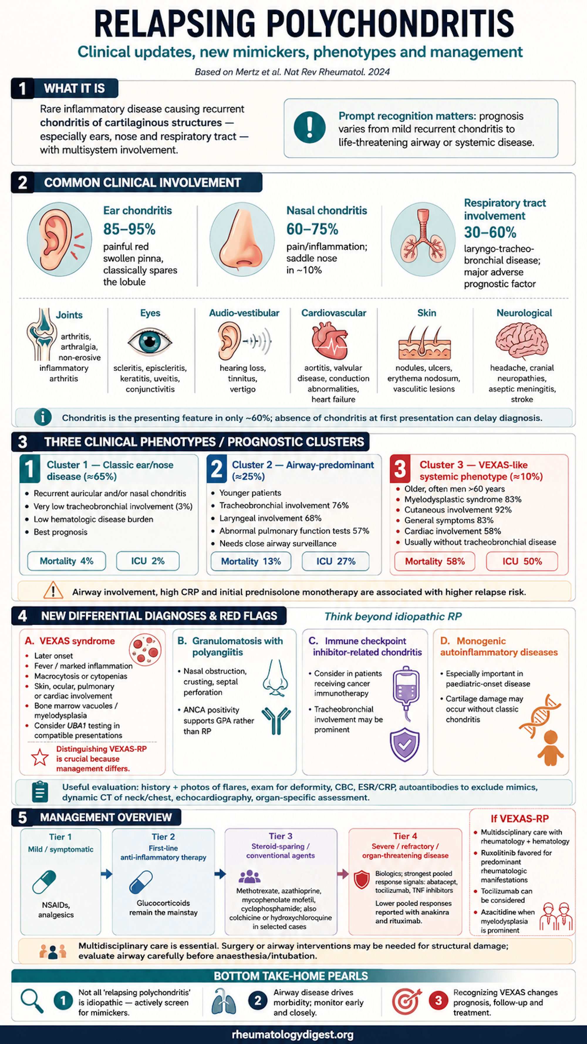
TL;DR: A tenth of what is called relapsing polychondritis is VEXAS, and what remains splits into three clusters whose mortality differs by more than an order of magnitude — which makes the diagnostic work, not the drug choice, where the clinical value now lies.
The clinical problem
A rare disease that is diagnosed clinically, has no confirmatory test, and — since 2020 — has a mimic that changes everything.
Relapsing polychondritis (RP) is a rare inflammatory disorder producing recurrent inflammation of cartilaginous structures, chiefly ears, nose and respiratory tract, accompanied by a broad range of systemic features. Estimated prevalence is 4.5–25 per million, incidence 0.71–3.5 per million per year, with the largest dataset being 256 cases across 124 million patient-years in Hungarian billing records (2002–2013). These figures should be read cautiously: rare and protean diseases are systematically underdiagnosed, and administrative databases depend on diagnosis codes with their own misclassification. Peak incidence falls in the fourth to sixth decades, cases occur at all ages including children, and most series show no clear sex predominance.
Four features make RP disproportionately difficult relative to its rarity:
- Chondritis — the defining feature — is absent at presentation in 40% of patients. Later onset of chondritis during the disease course is a major driver of diagnostic delay. The clinician is frequently asked to recognise RP before its cardinal sign has appeared.
- There is no diagnostic test. No autoantibody, no biomarker, no imaging finding is specific. Diagnosis rests on clinical pattern plus exclusion.
- Prognosis is genuinely bimodal. Standardised mortality ratio versus the general population is 2.16 (95% CI 1.24–3.51) in a UK population-based cohort, with respiratory disease, cardiac conditions and cancer the leading causes of death; in a Chinese series of 295 patients, respiratory failure from airway obstruction dominated. Yet many patients have mild, self-limited chondritis.
- The differential has been rewritten. The 2020 description of VEXAS syndrome retrospectively reassigned an entire historical subgroup of “RP with haematological abnormalities” to a different disease with different biology, different treatment and radically worse outcomes.
The central clinical proposition of this review: RP is not one disease with variable severity but a clustered phenotype with distinct prognostic trajectories, and a substantial share of what was called RP is actually something else.
Pathogenesis — genetic predisposition without a clear mechanism
Pathogenesis remains poorly defined. The current picture:
Genetic associations:
- HLA-DRB1*16:02, HLA-DQB1*05:02, HLA-B*67:01 — in linkage disequilibrium with each other.
- A whole-exome case–control analysis identified ultra-rare damaging germline variants in DCBLD2, with four specific variants in RP patients: missense G261V and I514F, stop-gain L250X, and frameshift Q435fs. These likely cause loss of function, and DCBLD2 appears to contribute to a complex, polygenic process rather than a monogenic form.
- Variation within the TNF pathway was also potentially associated.
- None of these alleles are tested in routine practice — a point the authors make explicitly, and which distinguishes them sharply from the actionable somatic genetics of VEXAS.
Immunology. Transgenic mice expressing HLA-DQ8 on a class-II-null background develop auricular chondritis and polyarthritis resembling human RP. Candidate autoantigens include type II collagen, matrilin 1, and COMP, though human data are inconsistent. Infiltrating T cells release IFNγ, IL-17 and TNF; experimentally, T cell responses against type II collagen residues 261–273 may have a specific role.
Environmental triggers: cartilage trauma including ear pinna piercing, and, notably, PD1 blockade with nivolumab.
The clinical spectrum — what to examine and what it means
Chondritis by site:
| Site | Frequency | Diagnostic pearls |
|---|---|---|
| Ear | 85–95% over disease course | Painful, red, swollen pinna sparing the lobule (non-cartilaginous) — the single most useful discriminating sign. Episodes last days to weeks, often resolve spontaneously. Episodes <24 h in untreated patients are unlikely to be RP. Deformity (cauliflower ear, thickening, calcification, floppy ears) in ~15% |
| Nose | 60–75% | Pain at the junction of nasal bone and cartilage, not the tip. Saddle nose in ~10%, insidious and usually painless |
| Respiratory | 30–60% | Major adverse prognostic factor. Hoarseness, dyspnoea, anterior neck pain, asthma-like presentation, stridor. Can occur in isolation, particularly lower airway |
| Costochondral / sternal | 20–40% | Chest pain, tenderness, swelling; rarely local deformity |
Two negative signs that earn their keep:
- Lobule sparing distinguishes ear chondritis from dermo-hypodermal infection. Transient, non-painful vasomotor redness lasting minutes to hours is a benign differential.
- Nasal obstruction, crusting and septal perforation point away from RP and towards GPA.
The GPA airway distinction — a genuinely practical CT discriminator:
| Feature | GPA | Relapsing polychondritis |
|---|---|---|
| Stenosis location | Mostly subglottic | Mostly tracheal |
| Morphology | Circumferential | Anterior, calcified, often extending to bronchi |
| Supporting features | Excavated lung nodules, chronic sinusitis, renal involvement | — |
Long-term airway sequelae include tracheobronchomalacia and dynamic collapse of the tracheobronchial tree. New or worsening upper airway symptoms warrant urgent evaluation. Patients with respiratory involvement have more costochondritis and pulmonary infection, and less ocular and auricular chondritis — an inverse relationship that recurs in the cluster analysis.
Systemic manifestations. Present at onset in 40% of new-onset RP; 20–30% have another associated autoimmune or inflammatory condition — the review lists MDS, lymphoma, Hashimoto thyroiditis, type 1 diabetes, PBC, myasthenia, SLE, Sjögren, APS, MCTD, RA, spondyloarthritis, Behçet and IBD.
- Joints (50–85%): migratory or additive, non-erosive, seronegative polyarthritis resembling seronegative RA or spondyloarthropathy. CRP is often normal even with demonstrable musculoskeletal involvement. Axial involvement resembles axSpA but characteristically lacks sacroiliitis on MRI.
- Eyes (50–70%; ~20% at presentation): episcleritis and scleritis each in about a third. Episcleritis recurs in parallel with systemic activity; scleritis is sight-threatening, with scleral thinning, necrosis and perforation. Uveitis in 23%, anterior far commoner than posterior (10.7% vs 2.6%). Ophthalmic evaluation is advised even in asymptomatic patients.
- Audio-vestibular (20–50%): sudden SNHL (glucocorticoids first-line, urgency matters for recovery), progressive SNHL (bilateral but asymmetric, needs serial audiometry), and vestibular dysfunction.
- Cardiovascular (5–10%): predominates in men over 60, particularly with VEXAS. Independent risk factors: age, CNS involvement, neutrophil-to-lymphocyte ratio >6.41, disease duration >4 years. Predominantly associated with airway involvement. Commonest valvulopathy is aortic insufficiency from annular dilation with ascending aortic ectasia; aortic involvement (aortitis, aneurysm, ectasia, dissection) in 5–10%, thoracic aorta most affected, with Takayasu-like stenoses or dilatation possible in other large trunks.
- Skin (20–40%, but see caveat): subcutaneous nodules, neutrophilic dermatoses including Sweet syndrome (may respond to colchicine or dapsone), cutaneous vasculitis (mostly leukocytoclastic on biopsy). The authors flag that most reported cases were men over 60 with haematological manifestations — i.e. probably VEXAS.
- Neurological (rare): CNS » peripheral. Trigeminal (V) and facial (VII) cranial neuropathies commonest, then aseptic lymphocytic meningitis; rare seronegative limbic encephalitis (predominantly men, mean age 59), myelitis, stroke-like episodes.
- Renal — mostly a red flag, not a feature. Historical series predating ANCA testing almost certainly overestimated renal involvement through misdiagnosed GPA. Anti-MPO and anti-PR3 are typically absent in RP; their presence should redirect the diagnosis.
MAGIC syndrome (Mouth And Genital ulcers with Inflamed Cartilage): 13 of 96 patients in an NIH cohort met criteria; none had UBA1 mutations. Predominantly female (70%), median age at diagnosis ~40 — demographically the mirror image of VEXAS. Higher prevalence of mucocutaneous ulcers, inflammatory eye disease, Behçet-type skin lesions, VTE, aortitis, GI involvement and CNS disease; Raynaud’s emerged as a possible feature. A proposed definition — meeting both McAdam’s/Damiani criteria for RP and the International Criteria for Behçet’s Disease — showed high sensitivity and specificity and awaits prospective validation.
VEXAS syndrome — the reframing that dominates this review
The historical puzzle solved. A 2015 cluster analysis had already identified an RP subgroup characterised by haematological abnormalities: mainly men over 60, increased mortality, myelodysplastic syndrome. The 2020 description of VEXAS supplied the missing mechanism. In a 2021 NIH cohort, 7.6% of 92 RP patients carried UBA1 mutations.
Mechanism. VEXAS arises from clonal expansion of haematopoietic stem/progenitor cells bearing somatic UBA1 mutations. UBA1 sits on the X chromosome and encodes the E1 enzyme initiating almost all cellular ubiquitination. Two isoforms exist: UBA1a (long, nuclear) and UBA1b (shorter, starting at codon 41, lacking a nuclear localisation signal, therefore cytoplasmic). Most pathogenic mutations affect the p.Met41 start codon of UBA1b or its intronic splice site, generating an isoform with impaired catalytic activity. Free ubiquitin accumulates in monocyte cytoplasm; reduced proteasome activity and accumulation of unfolded proteins drive an inflammatory state. Peripheral blood shows raised IL-1β and IL-18 (inflammasome activation, myeloid dysregulation) plus enrichment of TNF and NF-κB signalling.
Phenotypic contrast — the practical discriminator:
| Feature | Idiopathic RP | VEXAS–RP |
|---|---|---|
| Age at diagnosis | 44 years | 66 years |
| CRP | 10 mg/L | 69 mg/L |
| Nose chondritis | 70% | — |
| Fever | — | 60% |
| Cutaneous involvement | Absent | 82% |
| Ocular involvement | — | 57% |
| Pulmonary infiltrates | Absent | 46% |
| Heart involvement | Absent | 11% |
| Blood involvement | Absent | Present |
| Ear chondritis, peripheral arthritis, VTE | No difference | No difference |
The clinically usable version: cutaneous, ocular, pulmonary or cardiac involvement, older age, fever, and a markedly elevated CRP should push you towards VEXAS. Ear chondritis and peripheral arthritis do not discriminate. Patients with a known UBA1 mutation show less severe cartilage involvement and less large-airway damage, often with an infiltrative inflammatory lung disease.
When to screen for UBA1:
- Strongly consider in men with RP onset after age 50 who have MCV >100 fL or platelets <200 × 10⁹/L, particularly with cytoplasmic vacuoles in myeloid and erythroid bone marrow progenitors (highly suggestive but not pathognomonic).
- Diagnosis is by direct Sanger sequencing of exon 3, covering the three commonest variants (p.Met41Leu, p.Met41Val, p.Met41Thr). Rarer exonic mutations, splice variants and small clones require other techniques.
- The age/sex rule is loosening. Postzygotic UBA1 variants have been found in non-haematopoietic tissue (nails), challenging myeloid-restricted mosaicism; VEXAS has been reported in a 23-year-old man; rare female cases occur with inherited (Turner) or acquired X monosomy. The authors’ position: screening may be indicated regardless of age and sex in a suggestive clinical context.
- A negative UBA1 does not close the question. RP with haematological abnormalities but no UBA1 mutation has been documented, suggesting other somatic mutations yet to be discovered.
The conceptual claim, which is the review’s sharpest: given its distinct pathophysiology, symptom range, prognosis and treatment, VEXAS should be regarded as a disease mimicker, not a subtype of RP.
One counterintuitive detail worth retaining: the presence of ear chondritis in VEXAS is associated with better prognosis, and patients with the p.Met41Val variant are less likely to develop ear chondritis. Reported chondritis frequency in VEXAS has also fallen with wider ascertainment — 64% originally, 53.3% in a Spanish cohort, and 0% in one later population carrying pathogenic UBA1 variants. The chondritis-centric view of VEXAS was an artefact of how the syndrome was first found.
Other mimics — and when to think of them
Immune checkpoint inhibitor–induced RP. Immune-related adverse events occur in ~10% of ICI-treated patients. Six cases of ICI-induced RP are reported (five nivolumab, one pembrolizumab). The pattern is instructive: manifestations resolve on ICI discontinuation and relapse on rechallenge — as close to a causality demonstration as case reports allow. Tracheobronchial involvement appears commoner than in idiopathic RP. EULAR points to consider recommend prompt rheumatology referral and aiming for ≤10 mg/day prednisone-equivalent so as not to compromise ICI efficacy.
Monogenic autoinflammatory mimics — the paediatric imperative. Paediatric-onset RP (mean age ~9 years, no sex predominance, ocular involvement and joint pain common, >50% with family history of autoimmunity but concomitant autoimmune disease uncommon) should trigger consideration of genetic testing after discussion with an autoinflammation specialist. The key discriminating principle: these conditions cause structural cartilage damage that is not caused by chondritis.
| Condition | Gene | Cartilage phenotype |
|---|---|---|
| NOMID/CINCA | NLRP3 (GoF) | Nasal deformity; urticaria-like rash, fever, articular and neurological involvement |
| Protein kinase Cδ deficiency | PRKCD | Nasal deformity; monogenic SLE, immunodeficiency |
| Hypomorphic RAG1 deficiency | RAG1 | Mid-face involvement mimicking saddle nose; granulomatous hyperinflammation |
| TAP/HLA class I deficiency | — | Mutilating necrotising granulomatous inflammation of skin, upper airways (sparing lungs), septal perforation and nasal cartilage destruction |
| SAVI | TMEM173 (STING, GoF) | Vasculopathy with ulceration progressing to auricular cartilage necrosis and nasal septal perforation |
| Familial chilblain lupus / Aicardi–Goutières | TREX1 | Cold-induced lesions; pinna and nose involvement reported |
| CDC42-associated | CDC42 | One case with severe tracheobronchial cartilage damage, autoinflammation, immunodeficiency |
Diagnosis — criteria that are all unvalidated
Three criteria sets exist: McAdam’s (3 of 5 clinical features), Damiani and Levine’s (3 features, or 1 feature plus histology, or 2 features plus response to glucocorticoids/dapsone), and Michet’s (2 major, or 1 major plus 2 minor). The authors’ assessment is blunt and should be taken seriously: none has been formally validated, all were developed in small cohorts, and none defines organ involvement precisely.
Practical diagnostic strategy:
- History: ask about prior undetected chondritic episodes. Since patients are usually not seen during acute flares, ask them to photograph subsequent flares — a low-tech recommendation with high yield in a disease where the sign is transient.
- Laboratory: ESR and CRP may be normal even during flares, especially in non-VEXAS RP. Complete blood count matters — it screens for the MDS/VEXAS axis. Rheumatoid factor, ANA and non-PR3/non-MPO ANCA may be non-specifically present; PR3 or MPO positivity should immediately redirect the diagnosis. Anti-type II collagen and anti-matrilin-1 have poor sensitivity, specificity and availability and are not recommended routinely. Serum COMP is a candidate activity marker but assays are not widely available and standardisation is lacking.
- Imaging: dynamic inspiratory–expiratory CT of neck and chest (± 2D/3D reconstruction) is the key modality — showing thickening, calcification, malacia, and the often-overlooked finding of air trapping — and simultaneously helps exclude GPA and assess the aorta. MRI distinguishes acute oedema from chronic fibrosis in tracheal and laryngeal disease. PFTs exclude asthma but track damage only in severe stages. ¹⁸F-FDG PET–CT can map extent including asymptomatic cartilage, but its role in monitoring therapeutic response is controversial. Transthoracic echocardiography should be performed regularly. Auricular ultrasonography is proposed but unvalidated and operator-dependent.
- Cartilage biopsy is not required. Histology shows perichondrial and matrix inflammatory infiltration, loss of cartilage architecture, chondrocyte destruction with pyknosis, and fibrosis or granulation with collagen deposition and fibroblast proliferation — but features vary by stage and histology alone is not diagnostic. Reserve it for strictly unilateral auricular symptoms, absent systemic involvement, or poor glucocorticoid response — that is, to exclude alternatives, including B-cell lymphoma, which can mimic RP.
Monitoring instruments:
- RPDAI — international consensus, 27 weighted elements. A 2021 validation in 170 patients showed positive correlation with CRP-to-albumin ratio, neutrophil-to-lymphocyte ratio and platelet-to-lymphocyte ratio; a 2023 study identified CRP-to-albumin ratio as an independent risk factor for disease activity (OR 4.42).
- Relapsing Polychondritis Damage Index — consensus-derived, 17 items, pending formal validation.
- Physician global assessment can quantify activity but shows minimal correlation with patient global assessment — a discordance worth naming in clinic.
The three prognostic clusters — the review’s organising insight
From a French retrospective cohort of 142 patients:
| Feature | Cluster 1 | Cluster 2 | Cluster 3 |
|---|---|---|---|
| Share of patients | 65% | ~25% | ~10% |
| Defining features | Recurrent ear and/or nose chondritis | Younger patients; tracheobronchial (76%) and laryngeal (68%) involvement; abnormal PFTs (57%) | Men >60; MDS 83%, general symptoms 83%, cutaneous 92%, cardiac 58%; no tracheobronchial involvement |
| Tracheobronchial | 3% | Dominant | Absent |
| Haematological | 2% MDS, 5% other | None | 83% MDS |
| Mortality | 4% | 13% | 58% |
| ICU admission | 2% | 27% | 50% |
| Remission | All long-lasting remissions occurred here | — | — |
Cluster 3 almost certainly corresponds to VEXAS–RP.
Two observations deserve emphasis. First, the fourteen-fold spread in mortality (4% → 58%) across clusters means that “relapsing polychondritis” as an undifferentiated label carries essentially no prognostic information. Second, airway involvement correlated negatively with auricular chondritis (77%) — the two severe phenotypes are largely non-overlapping, which is precisely why a patient with isolated airway disease and no ear involvement is both the most dangerous and the most easily missed.
Other prognostic factors: relapse risk is increased by tracheal involvement, high CRP, and initial prednisolone monotherapy. Pregnancy does not modify the disease course.
Treatment — entirely off-trial
The evidence base is the central problem: there are no RCTs. All recommendations derive from observational studies and case reports. Only French guidelines (Health Authority/FAI2R) and selected ReCONNET guidance exist; no formal international recommendations have been issued.
Glucocorticoids (first-line, not evidence-based; French recommendations):
| Manifestation | Regimen |
|---|---|
| Active ear/nose chondritis | 0.5–1 mg/kg/day prednisone-equivalent until improvement, then taper over 10 days |
| Joint manifestations | 15–20 mg/day (not exceeding 0.5 mg/kg/day) over a few days, then taper to minimum effective dose |
| Laryngeal/tracheobronchial | 0.5–1 mg/kg/day (max 60–70 mg) for ≥3 weeks, then taper aiming for ≤15 mg at 3 months and ≤10 mg at 6 months |
| Severe disease | Methylprednisolone 250–1000 mg/day for 1–3 days |
Real-world dependency is high: median prednisolone 12 mg/day in a 2023 UK series of 68 patients, and 10 mg/day (range 5–12.75) at first recurrence in a Japanese series of 34.
Conventional DMARDs and biologics — pooled response rates from a 2022 systematic review (31 studies, 177 patients, 247 treatment lines):
| Agent | Response rate | 95% CI |
|---|---|---|
| Abatacept | 72% | 42–95 |
| Tocilizumab | 66% | 49–82 |
| TNF inhibitors | 64% | 53–74 |
| Methotrexate | 56% | 37–73 |
| Anakinra | 47% | 26–68 |
| Rituximab | 43% | 20–68 |
The authors’ reading is that abatacept, tocilizumab and TNF inhibitors are potentially superior to methotrexate, and that anakinra and rituximab should be avoided as much less efficacious.
A statistical caution worth registering. These confidence intervals overlap almost completely — abatacept’s spans 42–95%, rituximab’s 20–68%. The point estimates create an apparent hierarchy that the intervals do not support. These are pooled uncontrolled response rates from heterogeneous case series with no comparator, no randomisation, and substantial channelling bias, since sicker or more refractory patients receive different agents. The conclusion that rituximab “should be avoided” is a reasonable clinical heuristic but rests on evidence far weaker than the language implies. Other data: azathioprine 38–100%, cyclophosphamide 25–100% — ranges so wide as to be uninformative. Biologic use was associated with improved survival in one 2023 study. Dapsone is used for minor chondritis and neutrophilic dermatosis but carries risks of methaemoglobinaemia, haemolytic anaemia and DRESS.
VEXAS–RP requires a different algorithm — this is where the diagnostic distinction earns its clinical value:
- Glucocorticoid dependency and conventional or biological DMARD use are comparable to idiopathic RP, but outcomes are not.
- Predominant rheumatological manifestations: JAK inhibitors, particularly ruxolitinib, as suggested first-line, followed by tocilizumab. Watch for cardiovascular and thromboembolic events, to which VEXAS patients are already predisposed.
- Predominant haematological manifestations linked to myelodysplasia: azacitidine may help in high steroid dependency, refractory disease, or severe underlying MDS; allogeneic stem cell transplantation if azacitidine is insufficient.
- Management requires joint haematology–rheumatology input.
ICI-associated RP: no therapeutic guidelines exist. Each case needs oncology–rheumatology discussion, balancing the risk of cancer progression on ICI withdrawal against the risk of tracheobronchial involvement, which is commoner in this phenotype.
Non-pharmacological management: laryngotracheal reconstruction, ear pinna and nasal reconstruction, simultaneous aortic valve and ascending aorta replacement, and cochlear implantation may be needed. Airway stenting is associated with worse prognosis. Ambulatory CPAP can help respiratory collapse. Critically: the structural integrity of the tracheobronchial tree must be assessed before any anaesthesia or intubation — a case report documents tracheomalacic collapse under general anaesthesia in undiagnosed RP.
Key takeaways
Diagnostic
- Examine the lobule. A painful, red, swollen pinna with a spared lobule is the most specific physical sign in the disease. Lobule involvement points to infection.
- Chondritis is absent at presentation in 40%. Do not require it to consider the diagnosis, and ask patients to photograph future flares — the sign is transient and you will not be there.
- Normal CRP does not exclude active RP, particularly non-VEXAS disease. Conversely, a CRP around 69 mg/L in an older man is a VEXAS signal.
- PR3 or MPO ANCA positivity should redirect you to GPA. Nasal crusting, obstruction, septal perforation, subglottic circumferential stenosis, lung nodules, sinusitis and renal disease all point the same way.
- Anti-collagen II and anti-matrilin-1 testing is not recommended. No autoantibody is specific for RP.
- A complete blood count is a diagnostic test in this disease — MCV >100 fL or platelets <200 × 10⁹/L in an older man is the entry point to UBA1 sequencing.
The three-question triage this review supports. When you diagnose RP, ask:
- Is there airway involvement? — the strongest adverse prognostic factor, may occur in isolation, and requires dynamic CT with expiratory phases. It also mandates airway assessment before any intubation.
- Is this VEXAS? — older man, fever, high CRP, cutaneous/ocular/pulmonary/cardiac involvement, macrocytosis, cytopenias. Sanger sequence UBA1 exon 3. Treatment becomes ruxolitinib/tocilizumab ± azacitidine, with haematology.
- Is this something else entirely? — ICI exposure (temporal relationship, tracheobronchial predilection), paediatric onset (monogenic autoinflammatory disease, structural cartilage damage without chondritis), or Behçet-overlap features (MAGIC syndrome).
Prognostic
- Cluster membership, not the diagnosis, carries the prognosis: 4% mortality (ear/nose disease, 65% of patients), 13% (airway disease, 25%), 58% (VEXAS-like, 10%).
- Airway and auricular phenotypes are largely mutually exclusive — the absence of ear involvement should raise rather than lower concern.
- Overall SMR is 2.16, with respiratory, cardiac and malignant causes of death predominating.
Therapeutic
- No RCT has ever been conducted in RP. Every treatment recommendation, including glucocorticoid dosing, is expert opinion or uncontrolled observation.
- Pooled biologic response rates favour abatacept, tocilizumab and TNF inhibitors over methotrexate, with anakinra and rituximab apparently weaker — but the confidence intervals overlap almost entirely and channelling bias is unaddressed.
- The diagnostic distinction changes the drug. Idiopathic RP → glucocorticoids plus conventional/biologic DMARDs. VEXAS–RP → ruxolitinib, tocilizumab, azacitidine, allogeneic transplant.
- Airway stenting worsens prognosis; consider CPAP for collapse; assess the trachea before anaesthesia.
Where the field is weakest. The review’s research agenda: true incidence and prevalence, disease-specific quality-of-life measurement, validation of diagnostic criteria and of the Damage Index, the role of PET–CT and ultrasound in monitoring, novel biomarkers, and — above all — randomised controlled trials.
Final take
The lasting message of this review is subtractive rather than additive. It does not offer a new treatment or a new biomarker; it removes a substantial population from the diagnosis of relapsing polychondritis and reassigns it to VEXAS, and it partitions what remains into three groups whose mortality differs by more than an order of magnitude. For the practising rheumatologist, that translates into a short and specific set of actions: examine the lobule, image the airway dynamically, check the blood count, sequence UBA1 exon 3 when the pattern fits, and ask about checkpoint inhibitors. The therapeutic literature remains thin enough that the diagnostic work is where most of the clinical value lies — which is an unusual but honest position for a review to take.