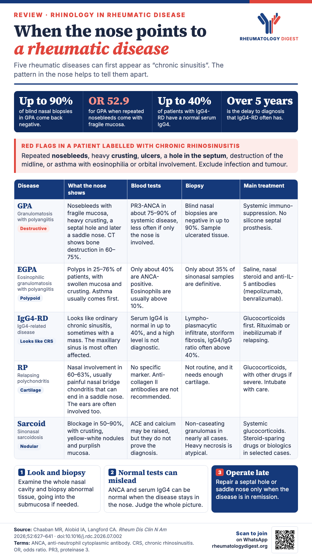
TL;DR: Chronic rhinosinusitis that bleeds, crusts heavily, ulcerates or destroys the septum is a red flag for a systemic rheumatic disease, and the pattern in the nose (destructive, polypoid, cartilage-based or nodular) points to which one, so examine the whole nasal cavity, biopsy visibly abnormal tissue, and do not let a negative blood test close the question.
Many sinonasal diseases begin by looking like simple rhinitis or sinusitis, and that is how a serious systemic illness can go unrecognised for years. This review, written by two otolaryngologists and a rheumatologist, is about the cases in which the nose is the first sign of a rheumatic disease. It describes how to tell them apart from ordinary chronic rhinosinusitis, how each disease shows itself in the nose, and how to treat it. Its central message is that surgery plays a supporting role, and that control of the underlying disease decides the outcome.
Chronic rhinosinusitis as the reference point
Chronic rhinosinusitis (CRS) is diagnosed when at least two of four cardinal symptoms are present: nasal obstruction, nasal drainage, facial pain or pressure, and reduced or lost sense of smell. There must also be objective evidence of inflammation on nasal endoscopy or CT, and the symptoms must have lasted more than 12 weeks. About 10–12% of adults in the United States have CRS, and it is divided into CRS with nasal polyps and CRS without nasal polyps.
Most patients improve with guideline-directed medical treatment and, where indicated, endoscopic sinus surgery. A subset do not, and in them the sinonasal disease is a sign of a systemic inflammatory or immune-mediated process. The authors list the clinical red flags that should make the clinician reconsider the diagnosis:
- recurrent epistaxis
- excessive crusting
- mucosal ulceration
- septal perforation
- destructive changes of the midline
- associated systemic features, such as asthma with eosinophilia or involvement of the orbit
General principles of care
Care of CRS in a patient with a systemic inflammatory disease should be shared between otolaryngology and rheumatology at every stage. Local nasal treatment is supportive and preventive in every disease covered here. Its goals are to reduce crusting, to limit trauma to the mucosa, to keep the mucociliary clearance working, and to lower the risk of secondary infection. The core measures are high-volume saline irrigation, humidification and lubrication, and prompt treatment of suspected bacterial superinfection.
Systemic immunomodulatory treatment is the cornerstone of disease control, and it often improves the nasal symptoms as well. Relapse can still occur, and loss of smell may be incomplete or irreversible. Surgery is reserved for selected patients with complications, or with obstruction that persists despite medical treatment. It is an adjunct, not a definitive treatment, because lasting control depends on managing the underlying disease.
Granulomatosis with polyangiitis
GPA is a systemic vasculitis that favours the upper airways, lungs and kidneys, with granulomatous inflammation and vasculitis of small and medium-sized vessels. It affects about 3 per 100,000 people, men and women equally, most often between the ages of 40 and 60. Ear, nose and throat involvement is often the first manifestation, so otolaryngologists are frequently the first to see these patients. Early symptoms are non-specific and centred on the nose, and many patients are first diagnosed with uncomplicated inflammatory CRS.
How the nose looks
Most patients have sinonasal symptoms. The common ones are reduced or lost smell, nasal obstruction, recurrent epistaxis, mucosal crusting, septal perforation and saddle-nose deformity. Endoscopy can show anything from redness and crusting to ulceration and marked friability. Persistent inflammation can cause synechiae and progressive destruction of cartilage, leading to septal perforation and, in advanced disease, a saddle nose.
The authors single out one finding: recurrent epistaxis with haemorrhagic, fragile mucosa is highly predictive of GPA (odds ratio about 52.9). It should raise strong suspicion once trauma and intranasal drug use have been excluded.
Laboratory tests
The work-up should include a full blood count with differential, a metabolic panel with creatinine and liver function, ESR and CRP, and, as the paper stresses, a urinalysis to look for kidney involvement.
- Cytoplasmic ANCA with anti-PR3 antibodies is found in about 75–90% of patients with systemic disease.
- Perinuclear ANCA with anti-MPO antibodies is found in 5–20%.
- ANCA may be negative in up to 20% of patients overall.
- ANCA is positive less often when the disease is confined to the sinonasal tract, with reported positivity of up to 60%.
Imaging
CT shows a mixture of active inflammation and accumulated damage to tissue and bone. The most common findings are mucosal thickening (about 85–90% of patients), bone destruction (60–75%), septal erosion or perforation (about 60%), and osteitis with new bone formation. The inflammatory changes in the mucosa can improve with treatment, but the bony changes often persist or progress. They therefore reflect the long-term burden of disease and not only active mucosal inflammation.
Biopsy
The classic histological triad is necrotising vasculitis, granulomatous inflammation and geographic necrosis. In practice, upper airway biopsies are often non-diagnostic, and blind nasal biopsies are negative in up to 90% of cases. The yield rises when the biopsy targets ulcerated, friable or necrotic areas and includes the submucosa. The supporting features (small-vessel vasculitis, multinucleated giant cells, granulomas, neutrophilic microabscesses and focal necrosis) appear together in only 16% of surgical biopsies. When the nose is unrewarding, tissue from another involved organ, such as the lung or kidney, usually gives a better yield.
Treatment
Many sinonasal symptoms improve once the systemic disease is controlled, but loss of smell may be incomplete or permanent, and many patients keep some congestion and crusting because of mucosal damage. GPA can be life-threatening, and systemic immunosuppression is the cornerstone: induction therapy to reach remission, with the drug chosen according to the organs involved and the severity, followed by maintenance therapy.
Surgery is reserved for complications or obstruction that does not respond to treatment. Septal perforations are usually managed conservatively. Silicone septal prostheses can be placed under local anaesthesia, but they are often poorly tolerated and may increase the risk of infection in GPA, so they are not recommended.
Eosinophilic granulomatosis with polyangiitis
EGPA, formerly called Churg–Strauss syndrome, affects 12–15 people per million. The mean age at diagnosis is 40–50 years, and men and women are affected equally. It typically moves through three overlapping stages: a prodrome of asthma and/or allergic CRS, then peripheral eosinophilia with eosinophilic infiltration of tissues, and finally systemic vasculitis. In most patients the CRS symptoms come several years before the vasculitis.
How the nose looks
Unlike GPA, sinonasal disease in EGPA is not destructive. Nasal polyps are particularly common, in about 25–76% of patients, and are often accompanied by reduced or lost smell. Examination usually resembles inflammatory CRS, with mucosal oedema, turbinate hypertrophy, crusting and polyps.
Laboratory tests
Although EGPA belongs to the ANCA-associated vasculitides, only about 40% of patients are ANCA-positive, most often with pANCA and anti-MPO antibodies. The two groups differ clinically: ANCA-positive patients more often have ear, nose and throat and kidney involvement, and ANCA-negative patients more often have cardiovascular complications. Other common findings are marked peripheral eosinophilia, typically above 10% on the blood count, and a raised ESR and CRP.
Imaging and biopsy
CT usually shows mucosal thickening and sinus opacification with no distinctive destructive pattern, often alongside nasal polyps. CT is not needed to make the diagnosis, but it helps to gauge the burden of CRS and to re-assess patients whose symptoms are atypical or out of proportion. Because EGPA is usually non-destructive, erosive or invasive sinonasal findings should prompt a search for another diagnosis.
The classic definition is tissue eosinophilia, necrotising vasculitis and eosinophil-rich extravascular granulomas, but these are often absent from sinonasal specimens. Only about 35% show definitive findings. Eosinophil-rich inflammation in nasal polyps is common but non-specific, because it overlaps with CRS with nasal polyps from other causes.
Treatment
Management of the nose in EGPA is mostly adjunctive, aimed at relieving symptoms and reducing airway inflammation. Local treatment is high-volume saline irrigation and topical intranasal glucocorticoids. The anti-IL-5 antibodies mepolizumab and benralizumab have been shown to improve CRS symptoms, including nasal obstruction and polyp burden, and to control asthma. EGPA can be life-threatening, especially with cardiac involvement, so the choice of systemic therapy should follow the pattern of organ involvement, the severity and the prognostic risk, for example by the Five-Factor Score.
IgG4-related disease
IgG4-RD was first described in 2003 as a cause of autoimmune pancreatitis. It is now recognised as a multisystem fibroinflammatory disorder that can involve almost any organ, including the paranasal sinuses. It usually presents in the sixth decade. The disease as a whole is more common in men, but this difference is less marked when the head and neck are involved.
How the nose looks
Sinonasal IgG4-RD causes symptoms that cannot be told apart clinically from CRS: nasal obstruction, rhinorrhoea, reduced smell and epistaxis. When lesions occur in isolation they can behave aggressively and destroy tissue progressively. The maxillary sinus is the most frequently affected site. Symptoms are often prolonged and resistant to treatment, and diagnostic delays frequently exceed 5 years. Endoscopy closely resembles other forms of inflammatory CRS, with diffuse mucosal oedema, turbinate hypertrophy, polypoid change or frank polyposis. In advanced or destructive cases, turbinate erosion or atrophy may be seen.
The 2020 revised comprehensive diagnostic criteria accommodate unusual sites such as the paranasal sinuses when laboratory or histological findings support the diagnosis. The authors add that the criteria are meant to support clinical judgement and not to replace it.
Laboratory tests
Serum IgG4 should be measured when IgG4-RD is suspected, but an elevated level is neither diagnostic nor a reliable measure of disease burden, and it may be normal in up to 40% of patients. Other possible abnormalities are raised IgG1 and IgE, low complement, and polyclonal hypergammaglobulinaemia.
Imaging
CT and MRI usually show non-specific inflammatory change (mucosal thickening or opacification) or mass-like lesions with remodelling or focal bone involvement. CT defines the extent of disease and the relationship of the lesion to the skull base and orbit. On MRI the lesions are typically isointense on T1, hypointense on T2 before contrast, and enhance homogeneously after contrast.
Biopsy
The characteristic triad is dense lymphoplasmacytic infiltrate, storiform fibrosis and obliterative phlebitis, although they are not always all present. Immunohistochemistry usually shows increased IgG4-positive plasma cells and a raised IgG4/IgG plasma cell ratio, commonly above 40%. Other inflammatory sinonasal conditions can also show plasma cell–rich inflammation, so the pathology has to be read together with the clinical and laboratory picture, with attention to whether the sample was adequate.
Treatment
Medical treatment combines saline irrigation and intranasal glucocorticoids with systemic glucocorticoids as first-line therapy for induction of remission. For recurrent, refractory or glucocorticoid-dependent disease, B-cell–directed therapy (for example rituximab or inebilizumab) can induce and maintain remission. Surgery is most often done to obtain a biopsy, which the authors call essential. It may also be needed to debulk or decompress lesions that cause mass effect or involve the orbit or skull base, and in refractory or progressive disease alongside systemic therapy.
Relapsing polychondritis
RP is a rare immune-mediated disorder of recurrent inflammation and progressive destruction of cartilage and other proteoglycan-rich tissue. The ear is the most commonly affected site, followed by the nasal cartilage, while the larynx and tracheobronchial tree are involved less often. An RP phenotype can occur in VEXAS syndrome, which is associated with myelodysplastic syndrome and has implications for prognosis and management. RP usually presents in middle adulthood, with a peak between the fourth and sixth decades, and the sexes are about equally affected.
How the nose looks
Sinonasal involvement occurs in about 60–63% of patients. It usually takes the form of nasal bridge chondritis, which is painful inflammation of the cartilaginous framework, and episodes recur. Repeated inflammation destroys cartilage progressively, and in severe cases it causes functional impairment and a saddle-nose deformity. Nasal disease usually accompanies other cartilage involvement, most often chondritis of the ear and less often laryngeal or tracheobronchial disease. Early nasal symptoms are non-specific and add to diagnostic delay, particularly when other systemic manifestations are present.
Laboratory tests, imaging and biopsy
There are no disease-specific biomarkers. ESR and CRP are often raised in active disease but are not specific. Antibodies to cartilage components such as type II collagen or matrilin-1 have poor sensitivity and specificity and are not recommended for diagnosis. Laboratory testing is therefore most useful for judging inflammatory burden, monitoring activity and excluding alternatives.
Imaging is supportive. It helps to find organ involvement, define structural damage and exclude mimics. In nasal disease, CT may show irregularity or deformity of the septum and cartilage, or soft-tissue inflammation, and it also looks for infection, GPA or malignancy. When a biopsy is done, it shows degeneration of the cartilage matrix, which loses its normal basophilia and looks more eosinophilic, with inflammation where the cartilage meets the perichondrium. The infiltrate is mixed (lymphocytes, plasma cells, eosinophils and neutrophils, with neutrophils often more prominent early), and chronic disease shows fibrosis and irreversible cartilage loss. Biopsy is not routinely required, and its yield depends on obtaining enough cartilage.
Treatment
Nasal treatment is mainly supportive, with saline irrigation, humidification and treatment of secondary infection, while systemic therapy targets the underlying chondritis. Systemic immunosuppression is individualised to disease activity and severity, with the aims of controlling inflammation, preventing irreversible cartilage damage and managing systemic manifestations. Glucocorticoids remain the cornerstone, and conventional immunosuppressants or biologics are used in severe, recurrent or refractory disease. Surgery is reserved for severe structural complications, persistent obstruction, or significant functional or cosmetic deformity.
One practical warning concerns anaesthesia. If a procedure needs general anaesthesia, intubation must be approached cautiously because of the possibility of tracheal stenosis and of airway collapse from loss of cartilaginous support.
Sarcoidosis
Sarcoidosis is a systemic inflammatory disease of unknown cause with granulomatous inflammation that can affect any organ. The course varies widely: more than half of patients reach remission, and about 30% have a chronic or relapsing course. In the United States the incidence is 10–20 per 100,000, rising to as high as 40 per 100,000 among African Americans. Women most often present in the fifth to sixth decades and men in the fourth to fifth.
How the nose looks
Sinonasal involvement is uncommon but significant. Nasal obstruction is the most frequent symptom (50–90%), followed by crusting, rhinitis, epistaxis, reduced smell, nasal polyps and facial pain. Some patients present with features outside the sinonasal tract, such as salivary gland swelling, nasal deformity, cervical lymphadenopathy, nasolacrimal duct obstruction, hoarseness or cranial nerve deficits. On endoscopy, the characteristic findings are yellow-white nodules, violaceous discolouration of the mucosa and polyps. Extracranial manifestations should raise suspicion in patients whose symptoms persist or resist treatment.
Laboratory tests, imaging and biopsy
Laboratory findings are generally non-specific. They include raised ESR and CRP, raised angiotensin-converting enzyme (ACE), hypercalcaemia and hypercalciuria. They can support the suspicion but are not diagnostic. CT often shows a mixture of CRS-like mucosal disease and nodular mucosal lesions on the septum or turbinates, and sometimes bone remodelling, erosion or new bone formation.
The diagnostic hallmark is non-caseating granulomatous inflammation. Granulomas are present in nearly all cases, unlike in many other inflammatory sinonasal disorders, so their absence should prompt consideration of another diagnosis. A little necrosis can occur, but prominent necrosis is atypical and should raise concern for infection or another granulomatous disorder.
Treatment
Saline irrigation and intranasal glucocorticoids can reduce crusting and ease symptoms, but treatment of sinonasal sarcoidosis relies mainly on systemic glucocorticoids. For patients who need a glucocorticoid-sparing approach, or who have more severe, relapsing or refractory disease affecting other organs, conventional immunosuppressants may be used, and biologics in selected cases. Surgery is mainly for obtaining diagnostic tissue, dealing with structural complications, or relieving symptoms that do not respond to medical treatment. Endoscopic procedures may improve nasal airflow and local control.
The five diseases side by side
| Nasal pattern | Blood tests | Biopsy | Mainstay of nasal-side care | |
|---|---|---|---|---|
| GPA | Epistaxis, friable mucosa, crusting, septal perforation, saddle nose | PR3-ANCA in about 75–90% of systemic disease; ANCA positivity lower in disease confined to the nose | Blind biopsy negative in up to 90%; target abnormal tissue | Systemic immunosuppression; no silicone septal prosthesis |
| EGPA | Non-destructive; polyps (about 25–76%), oedema, crusting | ANCA-positive in about 40%; eosinophils above 10% | Definitive findings in about 35% of sinonasal specimens | Saline, topical steroid, anti-IL-5 antibody |
| IgG4-RD | Looks like CRS; masses, orbital extension possible; delay over 5 years | Serum IgG4 normal in up to 40% | Lymphoplasmacytic infiltrate, storiform fibrosis, obliterative phlebitis | Systemic glucocorticoids; rituximab or inebilizumab if refractory |
| RP | Painful nasal bridge chondritis, saddle nose; usually with ear involvement | No specific marker; anti-collagen II not recommended | Not routine; cartilage needed | Systemic therapy; caution at intubation |
| Sarcoidosis | Obstruction, crusting, yellow-white nodules, violaceous mucosa | ACE, calcium; non-specific | Non-caseating granulomas in nearly all | Systemic glucocorticoids |
Reconstruction: septal perforation and saddle nose
Septal perforation
Patients with a symptomatic perforation commonly have obstruction, crusting, recurrent epistaxis, discharge and whistling on breathing, and quality of life can decline progressively. A careful endoscopic examination should assess the perforation by size, shape and location, since these guide planning. The incisive canal serves as the landmark between anterior and posterior defects. Before any repair, a medical assessment must confirm that the disease is quiescent.
The choice of technique depends on the location of the defect and the structural support around it. Routine interposition grafts are discouraged because closure rates are similar and morbidity is higher, except when combined with a pedicled septal flap. Current practice relies on mucosal advancement and vascularised pedicled flaps, particularly from the lateral nasal wall, which reliably cover moderate and large defects. Very large or near-total perforations may need a regional flap such as a pericranial flap.
Saddle-nose deformity
Reconstruction must restore both the airway and the stability of the framework, usually with load-bearing cartilage or bone grafts and not with simple mucosal repair. Continuing immune-mediated injury, vasculitic ischaemia and fibrosis impair healing and graft integration, which raises the risk of resorption, flap failure and recurrence. Autologous cartilage is generally preferred because it is biocompatible and resists infection, although long-term success depends on good blood supply and sustained disease control.
Reconstruction should be done only during remission, since active disease raises the risk of tissue breakdown and necrosis. The authors recommend a stepwise approach: costal cartilage grafts for mild to moderate deformity, and osseocartilaginous rib grafts for severe cases. Rigidity, contour and durability take priority in compromised tissue, and endoscopic visualisation may help planning but does not replace robust structural support.
Practical points from the authors
The review closes with a short list of care points, which fit together as a working approach to the patient with “CRS that is not behaving”:
- Broaden the differential when red flags are present. CRS plus epistaxis, crusting, ulceration, septal perforation or destruction should make the clinician think of rheumatic disease, while excluding infection and neoplasm.
- Examine the whole nasal cavity and biopsy the abnormal tissue. Look for friability, ulceration, crusting, polyps and masses, and sample abnormal areas, going into the submucosa if needed, because blind biopsies are often non-diagnostic.
- A normal blood test does not exclude disease. ANCA and serum IgG4 can be normal in localised disease, so the diagnosis rests on correlating the clinical, laboratory and pathology findings.
- Use CT or MRI to look for destructive patterns. Osteitis, erosion, loss of the septum or turbinates, extension into the orbit, or mass effect should lead to a systemic work-up.
- Match the pattern to the disease. GPA is epistaxis, friable mucosa, crusting, perforation and saddle nose. EGPA is asthma, polyps and eosinophilia. IgG4-RD is refractory CRS with a mass, orbital or salivary gland disease. RP is painful chondritis, with or without ear or airway involvement. Sarcoidosis is lung disease, nodules or violaceous mucosa, or lupus pernio.
- Reconstruct late and structurally. Septal and saddle-nose repair needs an algorithmic approach, structural grafts and timing during remission.
What it adds up to
The authors conclude that sinonasal symptoms are common and are usually attributed to CRS, but in some patients they are an early sign of systemic inflammatory disease and may come days to years before other organs are involved. Red flags should prompt a broader differential and systemic evaluation. Management is mostly medical, with surgery kept for diagnosis, complications and reconstruction after disease control. Joint care by otolaryngology and rheumatology is, in their words, critical to guiding management and optimising outcome.
Disclosures noted in the source: the authors report none. They state that they used ChatGPT (OpenAI) to assist with language editing and take full responsibility for the content.