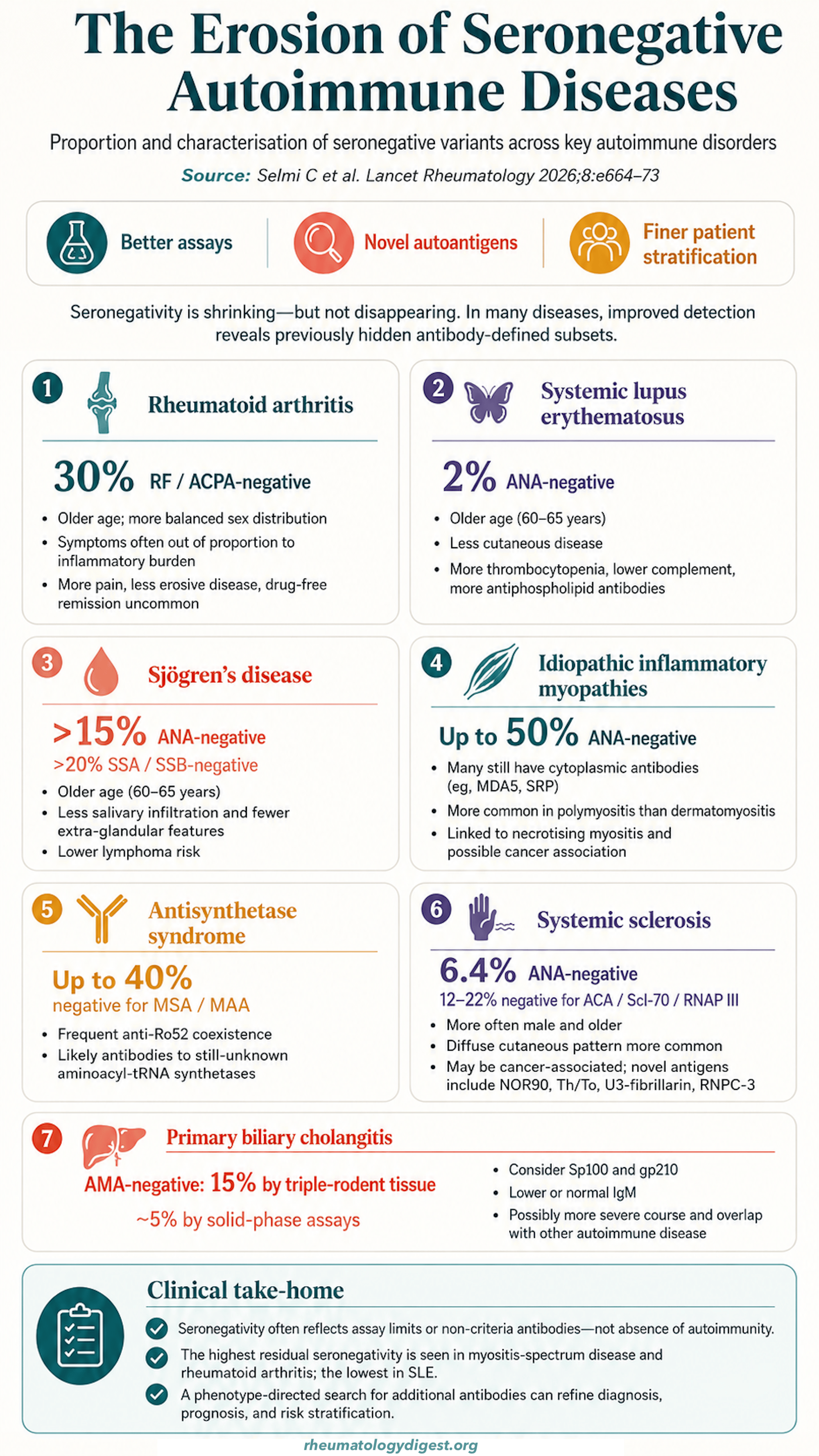
TL;DR: “Seronegative” conflates two opposite situations — an autoantibody the assay could not detect, versus a disease genuinely not driven by autoantibodies at all — and separating them changes everything: the first shrinks as assays improve (PBC seronegativity fell from ~15% to ~5%), while the second identifies myeloid/CD8-driven, MHC class I-associated disease that will not respond to B-cell depletion.
The Clinical Problem
“Seronegative” is one of the most consequential words in rheumatology, and one of the least examined. Applied to a patient, it changes the diagnostic label, the confidence of the diagnosis, the treatment offered, eligibility for trials, and — through all of these — the outcome. Yet the term conflates two entirely different situations:
- The patient has autoantibodies, but the assay used could not detect them, or the relevant specificity has not yet been discovered.
- The patient genuinely has no pathogenic autoantibody response, because the disease is driven by something else — innate immunity, CD8⁺ T cells, MHC class I biology.
These have opposite implications. The first is a technical problem with a technical solution; the second is a biological statement that should change how we conceive of the disease and what we treat it with. Historically we have not distinguished them, and the consequences are visible: seronegative rheumatoid arthritis is less well characterised than its seropositive counterpart, is systematically under-represented in randomised trials, and carries different disease severity, therapeutic response, residual pain burden and comorbidity profile.
The authors’ organising claim: autoimmune diseases showing B-cell-driven pathology and characteristic MHC class II associations are inevitably associated with autoantibody positivity — so within that group, seronegativity should shrink towards zero as assays improve. The interesting question then becomes: what is left over, and what does the leftover tell us?
How Much Seronegativity Is Really Out There
| Disease | Seronegativity rate | Phenotype of the seronegative patient |
|---|---|---|
| Rheumatoid arthritis | RF and ACPA negative in ~30% | More balanced sex distribution, older, symptoms disproportionate to inflammatory burden, higher pain burden (fibromyalgia and residual pain despite controlled inflammation), less erosive, drug-free remission seldom achievable |
| SLE | ANA-negative in only ~2% | Older (60–65 y), fewer cutaneous manifestations, more thrombocytopenia, lower complement, more antiphospholipid positivity |
| Sjögren’s disease | ANA-negative >15%; anti-Ro/La negative >20% | Older, less salivary gland infiltration, fewer extraglandular features, lower lymphoma risk (which tracks with RF and cryoglobulinaemia) |
| Idiopathic inflammatory myopathies | ANA-negative up to 50% | Partly an assay artefact — many have cytoplasmic antigens (MDA5, SRP) a nuclear-pattern ANA misses. Commoner in polymyositis, associated with necrotising myopathy, may be cancer-associated |
| Antisynthetase syndrome | MSA/MAA negative up to 40% | Frequent anti-Ro52 coexistence; antibodies to as-yet-unknown aminoacyl-tRNA synthetases |
| Systemic sclerosis | ANA-negative 6.4%; negative for the 3 classic specificities 12–22% | Commoner in men and at older age, more often diffuse than limited, may be cancer-associated. Novel antigens: NOR90, Th/To, U3-fibrillarin, RNPC-3 |
| Primary biliary cholangitis | AMA-negative 15% on triple rodent tissue, only 5% on solid-phase | Remainder often carry ANA to Sp100 or gp210; lower or normal IgM; possibly more severe course |
The Assay Is the Diagnosis — a Methodological Argument with Teeth
The authors make a point that deserves to be uncomfortable: classification criteria specify the antibody but not the method.
Anti-dsDNA illustrates it perfectly:
- The Farr radioimmunoassay is the notional gold standard — highly specific, but detects only high-affinity antibodies, with sensitivity around 65%.
- The Crithidia luciliae immunofluorescence test has excellent specificity but is semi-quantitative, operator-dependent and insensitive.
- ELISA and automated chemiluminescence achieve excellent sensitivity at the cost of specificity.
Since SLE classification criteria require “anti-dsDNA positivity” without naming a method, the same patient can be classified differently depending on which lab ran the test — and there is no guidance on which assay to choose for which purpose (high-probability diagnosis vs low-probability screening vs activity monitoring).
Parallel examples run through the paper:
- AMA in PBC: identification of the PDC-E2 subunit as the dominant antigen, plus recombinant-antigen solid-phase assays, cut seronegativity from ~15% to ~5%. Conversely, some patients are AMA-positive on HEp-2 cell-based assays but negative on PDC-E2 assays — implying further mitochondrial antigens remain undiscovered. Supporting this, antibodies to mitochondrial respiratory chain complex 1 have been described in inclusion body myositis.
- RF: the historical Waaler–Rose red-cell agglutination assay detected mostly IgM; ELISA performance depends on isotype, clonality and target (human Fc vs human IgG vs rabbit IgG).
- ACPA: anti-CCP2 IgG is standard, based on citrullinated synthetic peptides; anti-CCP3 incorporates multiple citrullinated epitopes with conformational structure, explicitly to widen the detectable antigen spectrum.
A telling asymmetry: 60–80% of SLE patients are anti-dsDNA positive, versus >90% of PBC patients being AMA positive. The authors read this gap as genuine pathophysiological heterogeneity — different autoantibodies produced by different B-cell populations at different differentiation stages, from CD20⁺ circulating B cells through to long-lived plasma cells. That has direct therapeutic relevance, since B-cell depletion spares long-lived plasma cells.
New Specificities Keep Eroding the Seronegative Fraction
- Antiphospholipid syndrome: criteria still rest on lupus anticoagulant plus IgG/IgM anticardiolipin or anti-β2GPI. “Seronegative APS” patients may carry IgA isotypes, or non-criteria specificities — antiphosphatidylserine, antiprothrombin, anti-domain I of β2GPI (these with promising clinical associations), or anti-annexin and antiphosphatidylethanolamine (less established).
- Rheumatoid arthritis: anti-CCP2 IgA in up to 10% of “seronegative” RA. Antibodies to other post-translational modifications — anti-carbamylated protein (6–20%), anti-acetylated protein (1–40%), anti-MDA-acetaldehyde protein (up to 30%) of seronegative RA. Caveat: these cross-react with ACPA, and their independent clinical relevance is unestablished.
Autoantigenomics — and the Awkward Finding It Produced
“Autoantigenomics” describes unbiased, high-throughput discovery of an individual’s full autoantibody repertoire, using protein microarrays and immunoprecipitation coupled with mass spectrometry. In SLE this has revealed a remarkably broad autoantibody spectrum with clinical correlations. Protein microarrays have a technical limitation — sensitivity to antigen conformation (native versus post-translationally modified) — which immunoprecipitation-MS largely avoids.
The awkward finding: healthy individuals share a substantial common autoantibody repertoire. This simultaneously complicates the interpretation of serological breadth in disease and creates a genuinely new research question — how to distinguish physiological or even protective autoimmunity from pathogenic autoantibody responses.
The Conceptual Map Worth Teaching
The paper’s central framework divides the space into overlapping domains:
- B-cell-driven autoimmune diseases (the core): autoantibodies as cornerstone of diagnosis, stratification and prognosis; benefit from B-cell depletion; usually MHC class II associations. Here autoantibody discovery should be pursued — RA, connective tissue diseases, ANCA vasculitis, PBC.
- Genuine seronegative diseases (limited value in autoantibody research): spondyloarthritis, Behçet’s disease, large vessel vasculitis, MHC-I-opathies, autoinflammatory RA. No detectable autoantibody, other immune mechanisms (myeloid, CD8⁺ T cells, NK cells), poor response to B-cell depletion, and MHC class I associations.
- Typically seronegative diseases with possible autoantibodies (poor clinical relevance): inflammatory bowel disease, primary sclerosing cholangitis.
- Post-infectious states (context-dependent): RF in endocarditis (bystander) versus HCV cryoglobulinaemia (disease-driving); parvovirus B19 and EBV as mimickers or triggers.
- Positive in healthy individuals (unknown, potentially protective): anti-DFS70, preclinical autoimmunity.
A specific warning worth quoting in teaching: ANCA can be detected in a relevant proportion of patients with active inflammatory bowel disease but has poor clinical relevance there. Autoantibody positivity does not make a disease autoimmune.
Stratification — Where Autoantibodies Have Already Rewritten a Specialty
Myositis is the paradigm. The field moved from the phenotype-based Bohan and Peter description to an autoantibody-based classification — antisynthetase, anti-MDA5, anti-TIF1-γ — reflecting distinct pathogenic pathways and stratifying patients by phenotype, histology, organ involvement and malignancy risk.
Concrete applications:
- Antisynthetase, anti-MDA5 and anti-PM/Scl antibodies mark elevated ILD risk, with differing radiological patterns and trajectories.
- Anti-Ro52 and anti-TRIM21 identify patients at higher risk of progressive fibrosing ILD.
- The contrast case: immune checkpoint inhibitor-associated myositis is autoantibody-negative and attributable to dysregulated CD8⁺ T-cell activation, with MHC class I — not class II — associations. This is offered as a clean example of genuinely seronegative autoimmunity. Nuance: transcriptomic profiling identifies a subset of ICI-myositis that is anti-TIF1-γ positive and resembles classical dermatomyositis.
The authors then raise treatable traits — imported from respiratory allergy, proposed in systemic sclerosis — as a possible successor to disease labels, noting that serum autoantibodies capture the heterogeneity of SSc better than cutaneous phenotype does. Whether autoantibodies should formally count as treatable traits, and how this applies to seronegative disease, is unresolved.
Autoantibodies as a Window on Pathogenesis
- SLE clustering: machine learning on longitudinal autoantibody profiles identifies clusters with distinct disease trajectories — and, importantly, changing trajectories track changing autoantibody profiles and titres. This challenges two dogmas at once: that autoantibodies are static, and that retesting is unnecessary.
- Anticentromere-related diseases: a proposed grouping spanning limited cutaneous SSc, Sjögren’s disease and PBC — sharing Raynaud’s, sicca, autoimmune epithelitis, B-cell activation. Profiling against MIS12C and CENP-C alongside CENP-A/B reveals major and minor specificity clusters.
- A unifying observation: most connective tissue disease autoantibodies target nucleic acid homeostasis and the transcription–translation machinery — dsDNA, topoisomerase, RNA polymerase, centromeres, aminoacyl-tRNA synthetases — and disrupted nucleic acid metabolism is itself a recognised pathogenic mechanism. Newer targets fit the pattern: telomere and telomerase-associated antigens (whose loss-of-function mutations cause pulmonary fibrosis) in SSc-ILD; RNA transcription factors; splicing factors (hnRNP); translation proteins (eIF2b).
- ACPA maturation in RA: ACPA are detectable in ~50% of asymptomatic individuals many years before clinical disease, and undergo immunological maturation — repertoire expansion, changes in Fc galactosylation and fucosylation, isotype switching — at the time of clinical synovitis onset. These changes are even more pronounced in those who develop RA-ILD. Whether anything analogous occurs in seronegative patients is unknown.
- Anti-MDA5 and COVID-19: the changed epidemiology of anti-MDA5 amyopathic dermatomyositis during the pandemic links a type I interferon signature shared by the autoimmune syndrome and SARS-CoV-2 infection, in which MDA5 is the principal antiviral sensor.
Cancer and Autoimmunity — a Bidirectional Serological Signal
- The landmark mechanistic observation: POLR3A (RNA polymerase III) mutations in tumours associate with anti-RNA polymerase III antibodies in cancer-associated systemic sclerosis — anticancer immunity spilling into autoimmunity.
- High-risk markers: anti-TIF1-γ and anti-NXP2 in myositis flag malignancy risk (with real-world heterogeneity).
- The counterintuitive one: in SSc, seronegativity itself — absent anticentromere, anti-topoisomerase I and anti-RNA polymerase III — carries substantially increased cancer risk, especially in the three years before or after disease onset. In these patients, rarer specificities such as anti-U3-RNP (fibrillarin) or anti-RNPC-3 (U11/U12-RNP) are often detectable. Here immunoprecipitation remains essential.
- At the other end: multiple concurrent autoantibodies correlate with reduced malignancy risk in both myositis and SSc — even in patients carrying a warning profile such as anti-TIF1-γ or anti-RNA polymerase III (e.g. coexisting anti-Sp4 or anti-CCAR1 with anti-TIF1-γ). This supports a role for autoimmunity in neoplastic clearance and raises the question of occult cancers as triggers.
“Is Less More?” — Three Cautions for Practice
- Complementarity. Autoantibodies must be integrated with clinical assessment, imaging and functional testing, in a multidisciplinary setting. Relying on serology alone risks assigning inappropriate diagnostic labels and producing equivocal management.
- Order the test knowing why. False positives and false negatives are both real; results cannot be interpreted without a working knowledge of the detection technique. The authors call for standardisation of protocols, reference ranges and reporting formats across laboratories.
- Tolerate ambiguity honestly. Inconclusive serology should lead to decisions only when evidence is robust — and should drive research into the less well-defined autoantibodies rather than premature clinical use.
Two illustrative endpoints sit close together: the reclassification of some patients with “seronegative RA” or “interstitial pneumonia with autoimmune features” as actually having antisynthetase syndrome (a win for discovery), and the finding of anti-annexin A4 antibodies in desquamative interstitial pneumonitis — a disease with an established environmental driver in smoking — suggesting autoimmune activation may be integral to pathogenesis rather than a byproduct.
The Research Agenda
Four domains, each with milestones achieved and work remaining:
- Method optimisation → define agreement between detection methods; establish reference standards; validate techniques for novel autoantibodies
- Spectrum expansion → continue autoantigen discovery and validate preliminary evidence
- Patient stratification → define new correlations; find autoantibodies predicting complications, progression and disease onset
- Trigger reveal → understand the link between triggers, autoantibody generation and disease development; identify individuals at risk
Key Takeaways
- “Seronegative” is usually a statement about the assay, not the patient. PBC seronegativity fell from ~15% to ~5% with recombinant PDC-E2 solid-phase assays; SLE is ANA-negative in only ~2%; a third of “seronegative” RA patients carry anti-CCP2 IgA or antibodies to carbamylated, acetylated or MDA-acetaldehyde-modified proteins. Before accepting the label, ask which assay was used and what it cannot see.
- Classification criteria name the antibody but not the method — and that matters. Farr RIA (~65% sensitivity, high specificity), Crithidia IF (specific, insensitive, operator-dependent) and ELISA/chemiluminescence (sensitive, less specific) will classify the same patient differently. There is currently no guidance on which to use for which clinical purpose.
- Some seronegativity is real, and it is biologically informative. Spondyloarthritis, Behçet’s, large vessel vasculitis, MHC-I-opathies and autoinflammatory RA cluster together: no autoantibody, myeloid/CD8⁺/NK-driven, MHC class I associations, and poor response to B-cell depletion. Recognising this predicts which patients will not benefit from rituximab far better than any serological test.
- Autoantibody positivity does not make a disease autoimmune. ANCA in active IBD is the cautionary example — detectable, and clinically near-meaningless in that setting. Context determines whether an autoantibody is a driver (HCV cryoglobulinaemia), a bystander (endocarditis-associated RF), an epiphenomenon, or possibly protective (anti-DFS70).
- Seronegative RA is a distinct clinical problem, not merely an unlabelled one. Older, more sex-balanced, less erosive, symptoms out of proportion to inflammation, greater residual pain and fibromyalgia burden, drug-free remission rarely achieved — and systematically under-represented in trials. Some of it may be genuinely autoinflammatory rather than autoimmune, which would also explain a share of treatment-refractory flares in seropositive disease.
- Myositis shows what autoantibody-based reclassification can achieve, and ICI-myositis shows its limits. The antisynthetase / anti-MDA5 / anti-TIF1-γ framework stratifies phenotype, ILD risk and malignancy risk; anti-Ro52 and anti-TRIM21 flag progressive fibrosing ILD. ICI-myositis, by contrast, is antibody-negative, CD8⁺-driven and MHC class I-associated — the same clinical syndrome arriving by a different immunological route.
- In systemic sclerosis, seronegativity is itself a red flag for cancer — absence of anticentromere, anti-topoisomerase I and anti-RNA polymerase III carries substantially increased malignancy risk, concentrated in the three years around disease onset, with anti-U3-RNP or anti-RNPC-3 often detectable on immunoprecipitation. Conversely, multiple coexisting autoantibodies correlate with lower cancer risk in both SSc and myositis.
- Autoantibody profiles are dynamic, not fixed. Longitudinal clustering in SLE shows trajectories changing in step with evolving autoantibody profiles and titres — undermining the dogmas of autoantibody permanence and of “test once, never retest.”
- Healthy people have autoantibodies too. Autoantigenomics has revealed a shared autoantibody repertoire in healthy individuals, which complicates any reflexive equation of serological breadth with disease and opens the question of what distinguishes physiological from pathogenic autoimmunity.
- Read this as a hypothesis, argued well, not as evidence. It is explicitly a Personal View by a group with substantial industry ties, synthesising a purposively selected literature. Its central proposition — that B-cell-driven, MHC class II-associated disease is inevitably seropositive — is a claim awaiting test, and the authors themselves state that the necessary first step is simply to define the boundary between genuinely seronegative and merely undetected-seropositive disease.