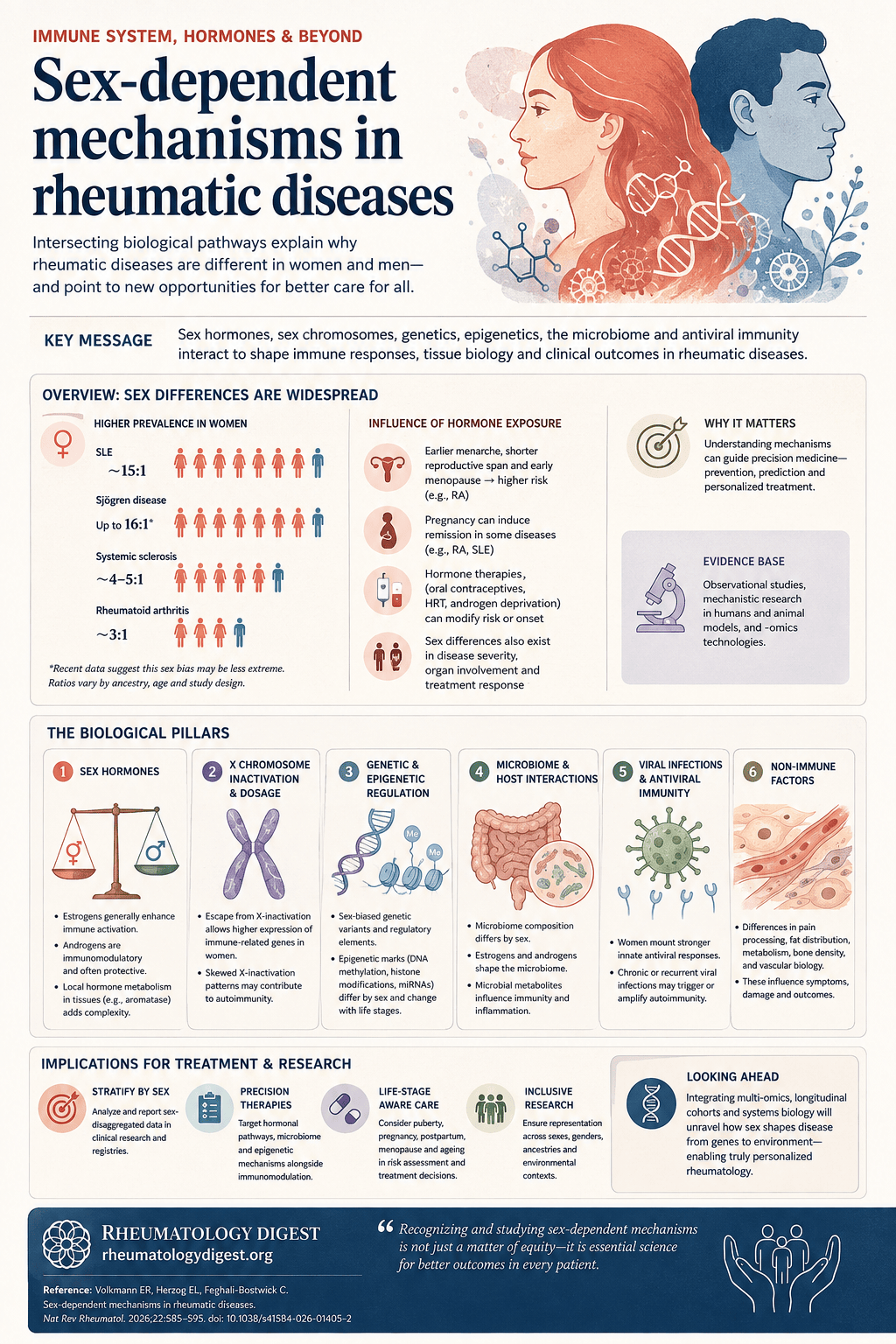
TL;DR: The female predominance of rheumatic disease is not one mechanism but three interlocking ones — sex hormones acting in disease-specific and sometimes opposite directions, X chromosome dosage operating independently of hormones, and a microbiome that reactivates oestrogen and feeds back into both.
The scale of the asymmetry
Roughly one in ten people live with an autoimmune disease, women are about twice as likely to be diagnosed as men, and autoimmune disease is the fourth leading cause of disability among women. In the autoimmune connective tissue diseases the skew is starker still, with female-to-male ratios approaching 15:1.
The observation is decades old. What has remained largely unexplained is why — and this review’s argument is that the answer is not simply “oestrogen”, but at least three intersecting axes: sex hormones, sex chromosome biology, and the microbiome, with antiviral immunity and epigenetic regulation layered on top.
A terminology point the authors make carefully, and which is worth preserving. The review uses ‘men’/‘male’ and ‘women’/‘female’ to match the language of the studies it cites, while acknowledging it is rarely clear how that information was collected or defined — whether by endocrine or chromosomal analysis, or by sex assigned at birth in medical records or registries. Sex and gender both exist on a spectrum, are not necessarily aligned, and a person with rheumatic disease might not identify as either a woman or a man. Practically: sex-related differences are widely reported across rheumatology, while gender-related determinants are far less consistently captured in clinical datasets — so the evidence base is uneven, and the discussion below reflects that unevenness rather than a considered choice to ignore gender.
What the epidemiology already tells us
| Disease | Female-to-male ratio | Hormonal association |
|---|---|---|
| SLE | approaching 15:1 | Most commonly diagnosed in premenopausal women; increased circulating oestrogen, reduced androgens |
| Primary Sjögren disease | historically up to 16:1 | Sex bias may vary across the lifespan; prevalence in both sexes tracks age-dependent testosterone and oestradiol trajectories |
| Systemic sclerosis | similar female predominance | Association with oestrogen; androgen deficiency implicated |
| Rheumatoid arthritis | 3:1 | Shorter reproductive span and early menopause linked to increased risk |
Two observations complicate the simple “oestrogen is bad” reading.
In SSc, androgen deficiency looks causally relevant. Disease onset has been reported in cisgender men after orchiectomy and androgen deprivation therapy for prostate cancer, and in transgender women receiving gender-affirming care involving orchiectomy or androgen suppression. That is about as close to a natural experiment as this field gets.
In RA, the direction of the hormonal signal depends on the compartment you measure. Epidemiology links shorter reproductive span and early menopause to higher risk, implying oestradiol is protective — yet RA synovial fluid shows increased oestrogen and reduced androgens, probably driven by local aromatase activity. Systemic and local hormonal environments are not the same thing.
In primary Sjögren disease, patients show reduced circulating DHEA, DHT and DHEA-S, plus impaired local androgen metabolism including decreased conversion of testosterone to DHT. Taken together, the authors propose that an imbalance between oestrogen and androgen signalling may be a shared mechanistic axis across these conditions — not a simple excess of one hormone.
Sex hormones: the machinery
Oestrogens differ by life stage — oestrone (E1) predominates after menopause, oestradiol (E2, 17β-oestradiol) during the reproductive years, and oestriol in pregnancy. They signal through nuclear receptors (ERα, ERβ), membrane-associated ERs, and G-protein-coupled receptors such as GPER. Androgens are produced primarily in the gonads, with the adrenals supplying precursors (DHEA, DHEAS, androstenedione, androstenediol). Testosterone can be converted to the more potent DHT or aromatised to oestrogens in many tissues — which is why “androgen level” and “oestrogen effect” are not independent variables.
Sex hormone receptors are expressed widely across immune cells and multiple tissues, which is what positions these pathways to regulate the sexual dimorphism seen in disease.
Adaptive immunity — and the central paradox
In B cells, oestradiol promotes survival, activation, class switching and somatic recombination. Individuals with two X chromosomes show oestrogen-dependent expansion of CD19⁺CD27⁺IgD⁻ class-switched memory B cells — highly efficient antibody producers, offering a plausible explanation for the broader and more pronounced humoral responses seen in women.
Then the picture inverts depending on the disease:
- In SLE, oestradiol further enhances B cell responses — disease-driving.
- In animal models of primary Sjögren disease, oestrogen deficiency promotes B cell accumulation, implying oestrogen is protective.
- In RA, postmenopausal oestrogen deficiency has been linked to reduced IgG sialylation in plasma cells, thereby increasing arthritis risk — again implying protection.
The same is true for T cells. Oestrogen has a critical role in thymic T cell development and dose-dependent effects on CD4⁺ differentiation: low-dose exogenous oestrogen promotes Th1 differentiation via ERα, whereas high-dose oestrogen favours expansion of immunosuppressive regulatory T (Treg) cells in both mice and humans. Since Treg cells are central to immune tolerance in RA, this may underpin oestrogen’s putative protective effect there. In SLE, by contrast, oestrogen appears to enhance T cell pathogenicity by increasing activation and survival.
One mechanism deserves separate mention because it operates on central tolerance itself: oestrogen epigenetically downregulates the autoimmune regulator (AIRE) gene in human and mouse thymic cells, impairing negative selection and promoting survival of autoreactive CD4⁺ helper T cells — a direct route from hormone to loss of tolerance in women with SLE.
In SSc the evidence is more indirect. Machine learning analyses of single-cell RNA-seq have shown that, relative to sex-matched healthy individuals, skin biopsies from women with SSc — but not men — are enriched for memory B cells and plasma cells alongside reductions in CD4⁺ memory T cells, though the relationship to hormones was not directly assessed.
A striking finding comes from SSc-ILD. Post-hoc analyses of bronchoalveolar lavage fluid from the SLS I and SLS II trials identified distinct, sex-biased molecular profiles that correlated with clinical phenotype: BALF from female participants — who responded better to lymphocyte-modulating therapy such as cyclophosphamide and mycophenolate — was enriched for inflammatory mediators including IL-12, IL-7 and G-CSF, while male samples were enriched for extracellular matrix remodelling proteins including MMP13 and TIMP1. In other words, the same diagnosis may be a more inflammatory disease in women and a more fibrotic one in men, which would explain differential treatment response without invoking hormones at all.
Innate immunity
Type I interferon-producing plasmacytoid dendritic cells (pDCs) recur throughout the sex-bias literature. In SLE and primary Sjögren disease, 17β-oestradiol markedly enhances TLR7- and TLR9-dependent IFNα production by pDCs stimulated with synthetic ligands or nucleic acid-containing immune complexes — direct mechanistic evidence linking oestrogen signalling to type I interferon, and a plausible account of why women produce more IFNα than men in both diseases.
TLR7 appears again in a spontaneous mouse model of SLE (dual knockout of Def6 and Swap70) that produces a female-predominant lupus-like syndrome. There, a distinct population of age-associated B cells (ABCs) — expressing conventional B cell markers together with T-bet and CD11c — preferentially accumulates in aged female mice, acquiring an interferon-stimulated gene signature and effector phenotype in a TLR7-dependent manner involving IRF5 and IRF8. ABCs have also been identified in women with SLE. The hypothesis worth noting: because this population is associated with ageing, declining oestrogen in older women might permit ABC expansion or activation — which would be oestrogen withdrawal, not oestrogen excess, driving disease.
In SSc skin, single-cell analyses show sexually dimorphic innate activation: samples from men showed enrichment of M0 and M1 macrophages with reductions in dendritic cells and M2-like macrophages, whereas samples from women showed enrichment of M1-like macrophages.
Non-immune cells and tissue remodelling
This is where SSc has been studied most, and where oestrogen’s effects are most clearly double-edged.
In dermal fibroblasts, oestradiol stimulates fibronectin production in primary cultures and human skin models, with increased dermal thickness, ECM deposition and TGFβ production. Mechanistically, dermal fibroblasts can generate their own oestradiol through IL-6-induced aromatase activation, converting androgens to oestrogens — a cell-autonomous, immune-independent feedforward loop. Consistent with a pathogenic role, selective ER modulators have been identified as potential SSc therapies using iPSC-derived dermal fibroblasts, organoid models and the repetitive bleomycin mouse model.
Yet the oestradiol metabolite 2-methoxyoestrogen (2-ME) does the opposite — reducing collagen deposition in mouse models of SSc-associated pulmonary complications, attenuating collagen I and CTGF expression in SSc dermal fibroblasts, and mitigating joint destruction, neutrophil infiltration and reactive oxygen species production in a mouse model of postmenopausal RA. A metabolite of the hormone antagonises the hormone.
Similar contradictions appear in synovium: 17β-oestradiol stimulates production of matrix-degrading enzymes and IL-1α in human fibroblast-like synoviocytes, but suppresses TAK1 activation and apoptosis in the same cells and in mice with collagen-induced arthritis.
The vascular dichotomy has direct clinical consequences. Agonistic anti-ERα antibodies have been detected in patients with SSc and correlate with disease activity and severity, supporting a pathogenic contribution. But short-term intravenous conjugated oestrogen induces rapid arterial dilatation in patients with SSc and in healthy individuals — within 15 minutes — improving endothelium-dependent vascular reactivity without affecting vascular smooth muscle; similar benefits are reported with oral conjugated oestrogen. Oestradiol is therefore both a profibrotic mediator and a macrovascular vasodilator in the same disease. The practical implication the authors draw: supplemental oestrogen might benefit patients with severe vascular complications at high risk of pulmonary arterial hypertension, while caution is warranted in those with lung fibrosis.
In primary Sjögren disease the evidence points the other way and is relatively consistent. Lacrimal glands from oestrogen-treated ovariectomised rabbits are protected from lymphocyte infiltration and glandular apoptosis; oestrogen deficiency induced by ovariectomy triggers lymphocytic inflammation followed by lacrimal acinar cell death in mouse models. A similar protective role has been described for DHT.
The X chromosome — dosage and inactivation
Beyond hormones, the X chromosome carries many immune-regulatory genes, and this is where some of the strongest human evidence sits.
X chromosome inactivation (XCI) transcriptionally silences one X in female cells during embryonic development, enforced by the long non-coding RNA XIST. But some genes escape XCI, producing higher expression in women — and disruption of XCI has been linked to autoimmune susceptibility.
Escape and XIST loss:
- In SLE, abnormal maintenance of XCI and aberrant expression of X-linked immunity genes in B cells — in both paediatric and adult patients — is thought to contribute to female predominance.
- In a subset of pDCs over-represented in the blood of women with SSc, the X-linked genes TLR7 and TLR8 escape XCI at increased frequency, accompanied by reduced XIST expression and heightened type I interferon activity.
- In mice with conditional deletion of Xist in B cells, spontaneous lupus with glomerulonephritis develops in a subset of animals (preprint).
- XIST RNA itself may be pathogenic in humans: elevated XIST levels in leukocytes from patients with SLE can act as a source of TLR7 ligands, stimulating pDC IFNα production in a TLR7-dependent manner.
Dosage — the aneuploidy evidence, which is the cleanest separation of chromosome from hormone available in humans:
| Karyotype | Finding |
|---|---|
| Triple X (47,XXX) | Occurs in ~1 in 344 patients with primary Sjögren disease and ~1 in 404 with SLE, versus ~1 in 1,000 in the general population |
| Triple X vs 46,XX | Primary Sjögren disease 2.9-fold more prevalent; SLE 2.5-fold more prevalent |
| Triple X vs 46,XY | Primary Sjögren disease 41-fold more prevalent; SLE 25-fold more prevalent |
| Klinefelter (47,XXY) | 14-fold increased risk of SLE versus 46,XY; several reports of adult-onset SSc over four decades |
| 47,XXY and 47,XXX | Increased prevalence of idiopathic inflammatory myopathy, particularly inclusion-body myositis |
Klinefelter syndrome is the decisive case: these individuals have congenital hypogonadism — low androgens and low oestrogens — yet a 14-fold higher SLE risk. The extra X, not the hormonal milieu, is doing the work.
One important negative: no enrichment of the 47,XXX karyotype has been observed among patients with RA, despite RA’s female predominance. Whatever drives the 3:1 ratio in RA, X dosage does not appear to be it — a useful reminder that “female-predominant” is not one mechanism.
Skewed XCI adds a further layer. In a cohort of 44 patients with RA, skewed XCI was observed in 31.5% of women with disease versus 17.4% of healthy women, and skewing has also been reported in larger cohorts of women with RA (n=110) and SSc (n=68).
A methodological caution the authors flag, and which matters for reading older literature. The Four Core Genotypes mouse model — which decouples sex chromosome complement from gonadal sex by relocating the testis-determining gene Sry — produced the influential observation that two X chromosomes alone are sufficient to promote a lupus-like phenotype through gonad-independent mechanisms. Subsequent work identified an unrecognised chromosomal translocation affecting Tlr7 gene dosage in some of these mouse lines, meaning certain earlier studies may have been confounded. The original untranslocated line preserves the model’s validity, but prior findings need careful interpretation.
Genetic and epigenetic regulation
Autoimmune-associated SNPs in SLE occur in genes involved in sex hormone signalling, immune pathways and epigenetic regulation — several in X-linked genes that may escape XCI, including TLR7, TLR8, NR3C4, IRAK1 and MECP2.
In RA, SNPs have been identified in the X-linked genes TIMP1 and IL9R, with the IL9R association reported specifically in men — consistent with unbuffered (hemizygous) expression of X-linked risk alleles. TIMP1 encodes an MMP inhibitor and IL-9R is involved in IL-9-mediated T cell development, supporting a role for X-linked variants in sex-biased joint degradation and T helper differentiation. To date, no X-linked genetic variant associations have been reported in primary Sjögren disease, SSc or IIM — an evidence gap rather than a negative finding.
X-linked microRNAs show sex-biased, disease-associated expression and are regulated by oestrogen. In female lupus-prone NZB/W F1 mice, miR-31, miR-155, miR-127 and miR-379 are differentially expressed in splenocytes compared with males — and are upregulated to comparable levels in oestrogen-treated, orchiectomised male lupus-prone mice, which is a neat demonstration that the hormone rather than the karyotype drives this particular signal.
A methylation loop ties this together: increased miR-21 and miR-148a in CD4⁺ T cells from patients with SLE and female lupus-prone mice is associated with reduced DNA methyltransferase expression, leading to DNA hypomethylation and increased expression of methylation-sensitive immune genes, including X-linked ones. Demethylation of genes on the inactive X contributes to overexpression of CD40L, CXCR3, OGT, miR-98, let-7f-2, miR-188-3p, miR-421 and miR-503 in CD4⁺ T cells from women with SLE compared with men — and the same overexpression can be induced in CD4⁺ T cells from healthy women by treating them with the DNMT inhibitor 5-azacytidine. That raises the possibility that drugs modulating DNA methylation could influence sex-biased immune responses.
The microbiome, and the loop it closes
Gut microbiome composition differs between male and female mice, and early-life microbial colonisation influences sex hormone levels with downstream effects on immune development.
The mechanism that makes this more than correlation: bacterial β-glucuronidase converts inactive conjugated oestrogens back into their active form, promoting reabsorption of oestrogen into the circulation. Gut microorganisms can likewise deconjugate glucuronidated androgens and modulate their metabolism. The microbiome is therefore not merely adjacent to sex hormone biology — it is part of the hormone’s regulatory circuit.
Evidence across models:
- In lupus-prone mice, gut dysbiosis tracks with disease progression, and microbial composition differs between female and male NZB×NZW F1 mice. Transferring caecal contents from male to female mice reduces proteinuria, attenuates kidney disease and improves survival.
- Translocation of Enterococcus gallinarum from gut to other organs triggers autoimmune features in lupus-prone mice; oral vancomycin or ampicillin reduces anti-dsDNA and anti-RNA antibody production and improves survival. Whether these processes differ between sexes has not been assessed.
- Molecular mimicry: several commensal bacteria encode orthologues of Ro60 and trigger anti-Ro60 autoantibodies and Ro60-autoreactive T cells in SLE. That study was conducted predominantly in samples from women, so extension to men requires further work.
- In SSc-associated pulmonary fibrosis models, low gut microbial diversity amplifies the profibrotic effects of oestrogen in female mice. A cross-sectional international study in patients with SSc found gut dysbiosis correlating with radiographic ILD severity and inflammatory markers.
Inflammation feeds back the other way: TNF, IL-1 and IL-6 can shift hormone levels — increasing oestrogen, decreasing androgens — and those hormonal changes in turn alter the intestinal microbiota. In primary Sjögren disease (predominantly women), disease was associated with enrichment of pro-inflammatory taxa and depletion of anti-inflammatory species.
The most elegant experiment ties chromosome to microbe directly: in the Four Core Genotypes model, XX mice but not XY mice show increased IgM-secreting B cells and plasma cells after immunisation with heat-killed Streptococcus pneumoniae. That sex-chromosome-dependent effect was abolished by antibiotic depletion of the gut microbiota and restored by reconstitution with short-chain fatty acid-producing bacteria — making microbial factors both sufficient and necessary for XX-dependent immune activation.
Viral infection and antiviral immunity
Chronic viral antigen exposure is the environmental axis. Associations between EBV and autoimmune rheumatic disease have been reported in SLE, SSc, RA and primary Sjögren disease, and both mouse and human data indicate women mount more robust antiviral responses than men, with EBV-infected women generating stronger IgG responses.
Mechanistically, single-cell sequencing with ATAC-seq and ChIP-seq in patients with SLE identified EBV-infected B cells in which EBV nuclear antigen 2 (EBNA2) binds transcriptional start sites and regulatory regions of genes involved in immune regulation and antigen presentation. This reprogramming drives expansion of autoreactive B cells with antigen-presenting features, enabling T cell activation and production of antibodies against neoantigens. The predominance of women in that study precludes sex-based comparison — so this is a mechanism that plausibly explains sex bias but has not yet been shown to.
Implications for treatment
Sexual dimorphism in treatment response is already visible in clinic: SLE, SSc and primary Sjögren disease tend to be more severe and treatment-refractory in men, whereas RA is often more severe in women.
Hormone-based therapy has been the obvious thing to try, and it has largely disappointed. DHEA in SLE showed minor and variable effects on disease activity across seven randomised controlled trials — with modest improvement in patient-reported quality of life but adverse effects including acne. Hormone replacement therapy and testosterone-based interventions in RA have yielded mixed results. The authors read these failures not as evidence that hormones don’t matter, but as evidence that we don’t yet understand which cell types and receptor pathways mediate the effects well enough to intervene bluntly.
Emerging directions under investigation: CRISPR–Cas9 approaches that could in principle correct skewed XCI or modulate X-linked gene expression; microbiome-targeted interventions including dietary modification, selective microbial depletion or phage therapy; and AI-based drug repurposing to identify targets related to sex-biased autoimmune consequences of antiviral immunity.
Two evidence gaps are named explicitly and both are clinically live:
- How gender-affirming hormone therapy influences autoimmune disease onset, progression and flares. Randomised trials are not feasible here, so carefully designed observational studies will be essential.
- How supplemental oestrogen affects autoimmune risk and progression, given the resurgence of hormone replacement therapy in postmenopausal women.
What remains unanswered
The review closes with the questions it cannot yet answer — worth reading as a research agenda:
- What are the effects of sex hormones across all the cell types involved in rheumatic disease?
- What are the effects of the various sex hormone metabolites?
- Do additional XCI-associated genes contribute to pathogenesis?
- Are distinct molecular or cellular pathways predominant by biological sex, and can these be selectively targeted?
- Which components of the microbiome are beneficial or harmful, and how are these influenced by sex?
The framing conclusion is a fair summary of where this leaves us: treatments for rheumatic disease are currently not sex-specific, despite substantial evidence that therapeutic responses differ between women and men. Variations in sex hormones, the microbiome, antiviral immunity, genetic architecture and epigenetic regulation probably all contribute to differences in prevalence, clinical manifestation and treatment outcome — and none of it currently reaches the prescription pad.
Reading this with the right caveats
This is a narrative review, not a systematic one, and much of the mechanistic evidence it assembles is animal or in vitro work — mouse models of SLE, ovariectomised rabbits, cultured fibroblasts, bleomycin models. Several of the most striking findings are single studies, at least two are cited as preprints not yet peer-reviewed (the Xist B cell deletion model and the RA myeloid interferon signature), and the Four Core Genotypes translocation problem is a live warning that this literature has already had to revise itself once.
The human evidence that is strongest — the aneuploidy prevalence data and the skewed XCI cohorts — rests on small numbers in places (44 patients with RA in the original skewing study; 68 women with SSc). And the SLS I/II BALF finding, which is arguably the most clinically suggestive result in the review, is a post-hoc analysis.
Two of the three authors declare consultancy relationships with pharmaceutical companies (Boehringer Ingelheim, GlaxoSmithKline, AbbVie, Bristol Myers Squibb, Merck), which is worth registering even though this is a mechanistic review rather than a trial report.
None of this undercuts the central argument, which is a claim about mechanism rather than about practice. But it does mean the appropriate response is to update how you think about sex differences in these diseases — not yet how you treat them.