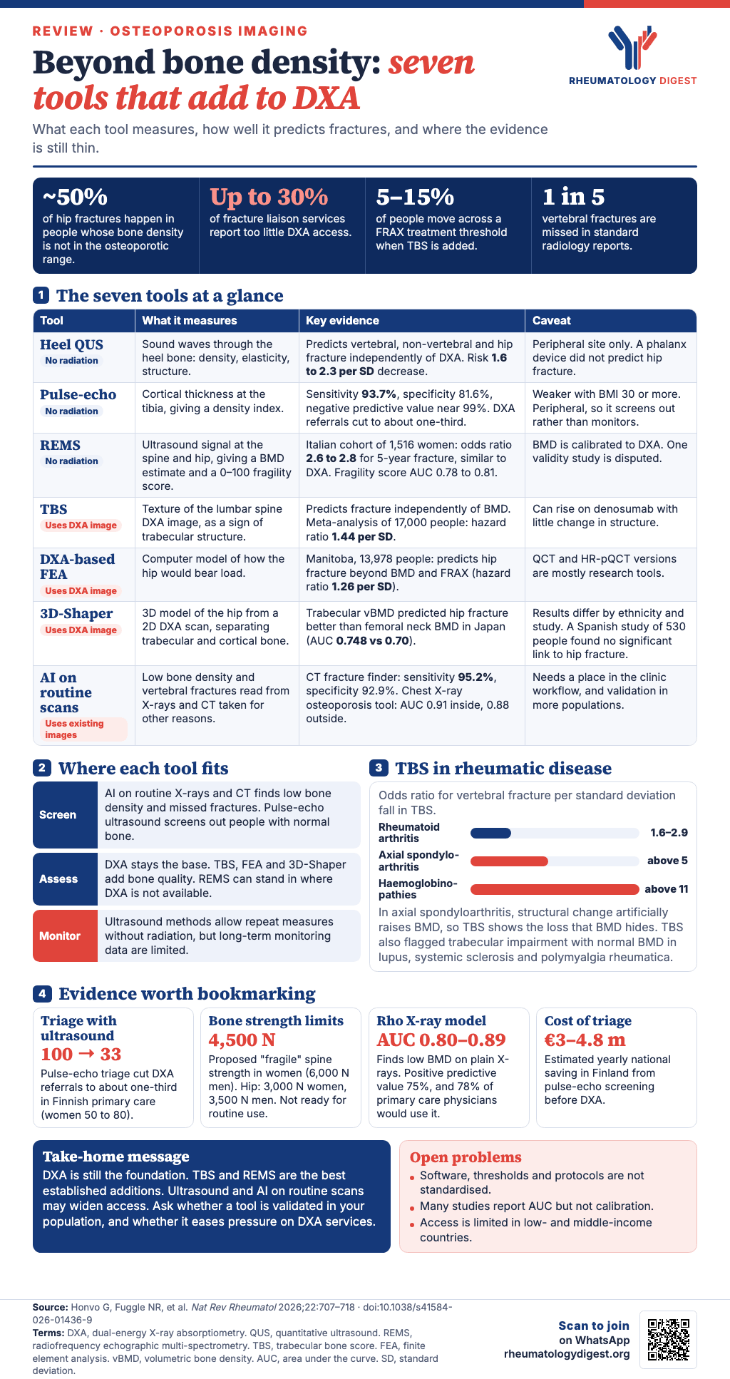
TL;DR: DXA bone density remains the foundation of osteoporosis assessment but captures only part of fracture risk and is hard to access in many places, and a set of single-component tools (ultrasound, trabecular bone score, finite element analysis, 3D shape modelling and AI on routine imaging) can add bone-quality information or reach people DXA misses, with trabecular bone score and REMS the best established and standardisation, validation and equity still unsettled.
Dual-energy X-ray absorptiometry (DXA) made it possible to define osteoporosis and moved the whole field forward, but it was never meant to carry the whole job of finding people at risk of fracture. This review, written under the WHO Collaborating Centre for the epidemiology of musculoskeletal health and ageing in Liège, surveys the newer tools that take a single measurement from one modality. These are different from multi-component tools such as FRAX, Garvan and QFracture, which combine clinical risk factors with a bone density result to estimate fracture probability over 5 or 10 years. Rheumatologists will find the review relevant, because its disease examples include rheumatoid arthritis, axial spondyloarthritis, lupus, systemic sclerosis, polymyalgia rheumatica and glucocorticoid-treated transplant recipients.
Why look beyond DXA
Osteoporosis is defined by a DXA T score of −2.5 or lower. Three problems follow from relying on that number alone:
- Many fractures happen outside the osteoporotic range. Around 50% of hip fractures occur in people whose bone density is not in the osteoporotic range, so areal bone mineral density (aBMD) by itself is not enough to guide treatment.
- Fracture risk depends on more than quantity of bone. Bone structure, microarchitecture and strength (the aspects of bone quality) also matter, and aBMD says little about how bone is distributed between the cortical and trabecular compartments.
- Access is uneven. DXA availability varies a great deal, operators are not always adequately trained, and reimbursement and financial limits persist. In established fracture liaison services, up to 30% report inadequate DXA access to meet clinical need. Calibration, operator-dependent error and inconsistent reporting add to the difficulty.
The review therefore covers three groups of tools: non-ionising techniques, image-analysis methods that estimate bone microarchitecture, and artificial intelligence (AI) applied to routine imaging. They can be used at different points in a patient’s journey, from opportunistic screening of people who already have a scan, to supplementing DXA at diagnosis, to repeated measurement during treatment.
Ultrasound, without radiation
Ultrasound avoids radiation risk, is portable and can reach people who are otherwise under-served.
Quantitative ultrasound at the heel
Quantitative ultrasound (QUS) measures how sound waves interact with bone. The best-studied site is the heel (calcaneum), where broadband ultrasound attenuation, speed of sound and a stiffness index derived from the two (calculated differently by each manufacturer) reflect density, elasticity and microarchitecture.
- Large meta-analyses show that lower QUS values predict vertebral, non-vertebral and hip fractures independently of DXA bone density, with risk estimates of about 1.6 to 2.3 per standard deviation decrease, similar to hip BMD. The associations survive adjustment for FRAX.
- Not every device and site performs equally. In more than 7,000 elderly women, heel devices predicted hip fracture, but a phalanx device did not.
- QUS has predicted fractures in chronic disease. In chronic kidney disease the hazard ratios are modest but consistent at all stages, and in type 1 and type 2 diabetes it often outperformed DXA, which suggests it picks up microarchitectural deterioration that DXA density does not.
Pulse-echo ultrasound
Pulse-echo ultrasound is a form of QUS that measures cortical thickness at the proximal tibia, which is a more stable peripheral site, and produces a density index that correlates with femoral neck and total hip BMD.
- In a large Finnish primary-care cohort of women aged 50 to 80, the multi-site density index identified DXA-defined osteoporosis with sensitivity of 93.7%, specificity of 81.6% and a negative predictive value approaching 99%. Used as a triage step, it cut DXA referrals to about one-third of participants. Sensitivity rose with age, and reproducibility was acceptable.
- A US multiethnic cohort found sensitivity near 80% and specificity close to 90% in both white and Hispanic women using the same cut-offs.
- Another study reported odds ratios above 3 per standard deviation decrease for DXA-defined hip osteoporosis, with sensitivity and specificity about 80–82%. The association was weaker with a BMI of 30 kg/m² or more, possibly because fat attenuates the signal. Triage pathways cut the need for DXA to about 27–31%.
- In an Austrian pilot of 33 older adults (mean age 82) with considerable comorbidity, clinical decisions for 90.9% of patients were made without DXA when pulse-echo ultrasound was used alongside FRAX, although specificity differed between men and women.
- A cost–utility analysis in Finnish women aged 65 to 85 found pulse-echo ultrasound, alone or followed by DXA, consistently cheaper than DXA-only pathways, with national savings estimated at 3 million to 4.8 million euros a year.
The review’s reading is that pulse-echo ultrasound is best seen as a way to screen out people with normal bone density and protect DXA capacity. Because it measures a peripheral site, it may be less helpful for axial bone health, and long-term monitoring with it may be limited.
Radiofrequency echographic multi-spectrometry (REMS)
REMS is a radiation-free, portable ultrasound method aimed at the axial skeleton: lumbar spine, total hip and femoral neck. It analyses the raw back-scattered radiofrequency signal, compares it with a reference library from people of matching age, sex, BMI and site, and reports two things.
- A BMD estimate with T and Z scores. This is calibrated against contemporaneous DXA, not measured directly as areal density.
- A fragility score from 0 to 100, based on the share of scan-line spectra that look more like a “fragile” model than a normal one. It aims to reflect microarchitecture-related fragility beyond aBMD, and it feeds 5-year fracture risk estimates built from manufacturer datasets.
What the studies show:
- REMS and DXA values correlate strongly in a large European multicentre study of postmenopausal women aged 30 to 90 (r = 0.94 for lumbar spine and 0.93 for femoral neck).
- In an Italian prospective cohort of 1,516 women, being classed as osteoporotic by REMS carried an odds ratio for 5-year fragility fracture of 2.6 at the lumbar spine and 2.8 at the femoral neck. Discrimination was comparable with DXA, and for vertebral fracture the lumbar spine AUC was 0.78 for both. For any fragility fracture at the lumbar spine, the AUC was 0.65 with REMS against 0.61 with DXA, and for fractures at sites other than the spine and hip 0.59 against 0.54.
- In a cohort of 1,989 women and men (248 incident fractures), the REMS fragility score predicted 5-year fracture in women (AUC 0.81 at the lumbar spine and 0.78 at the femoral neck) and men (0.78 and 0.81). The lumbar spine fragility score had an odds ratio of 9.2 for incident fragility fracture, and the femoral neck score an odds ratio of 6.4 for incident hip fracture. For comparison, the REMS T score had an odds ratio of 3.58 and the DXA T score 2.50 at the lumbar spine.
- In a cross-sectional comparison of 223 people with type 2 diabetes and 102 without, REMS gave lower spine and total hip BMD than DXA, labelled more people osteoporotic, and its BMD and fragility score both separated people with earlier major fractures from those without. These are cross-sectional data, and longitudinal confirmation is needed.
- In 30 kidney transplant recipients on long-term glucocorticoids, all fell into the high-risk classes, in keeping with what is known about these patients, though the study was small.
- Results from Brazil, Mexico, Japan, Colombia and Indonesia resembled those from Europe.
REMS can be used where DXA radiation is unacceptable (for example in pregnancy and children) and where people cannot travel to a DXA unit, such as those in domiciliary care or on an inpatient ward. It may also suit health facilities that cannot afford DXA infrastructure or a trained workforce, and modelling suggests REMS-based pathways could be cost-effective in Italy and the USA.
A live controversy. One study suggested that 91% of the variance in REMS femoral neck BMD can be explained by age, sex and BMI, and that BMD at the hip did not change before and after total hip replacement with the prosthesis in place. Later publications have questioned the technical acquisition and the statistics behind that study, and the matter is still debated. The review concludes that prospective 5-year data support REMS T scores as a feasible adjunct or alternative to DXA in some settings, and that the fragility score may add information on bone quality, but that further validation is needed.
Looking at bone quality on DXA and CT
BMD measures bone quantity. The tools below try to say something about quality.
Trabecular bone score
Trabecular bone score (TBS) is a texture analysis of the lumbar spine DXA image that indirectly reflects trabecular microarchitecture. It was calibrated against 3D features of cadaveric bone.
- Fracture prediction independent of BMD. Early studies showed lower TBS in people with vertebral or major osteoporotic fractures, with odds ratios for vertebral fracture of 1.3 to 2.0 per standard deviation decrease. In a Manitoba cohort of 29,000 women, hazard ratios for major osteoporotic fracture were 1.20 to 1.40 per standard deviation, independent of lumbar spine BMD. In more than 3,600 older men, AUCs were 0.59 for major osteoporotic fracture, 0.67 for hip fracture and 0.57 for clinical vertebral fracture. A meta-analysis of over 17,000 participants found a hazard ratio of 1.44 per standard deviation, and TBS predicts fracture in both sexes and across studies.
- Effect on FRAX. Adding TBS to FRAX in studies of 2,500 to 50,000 people increased the AUC only slightly, but it moved 5–15% of people across treatment thresholds, against 1.1–3.0% in the Manitoba BMD cohort. Reclassification was most marked in younger postmenopausal women, people with osteopenia and those with lower BMI, so TBS is most useful when BMD alone discriminates poorly.
- Improved algorithm. A soft-tissue correction, derived directly from the DXA image, addressed earlier limits in people with a higher BMI. In 1,362 postmenopausal women the odds ratio per standard deviation rose from 1.43 to 1.54, and a second study of 1,632 women found similar gains across vertebral combinations.
- On treatment. In two registry studies of women treated with bisphosphonates (4,838 and 5,536), TBS still predicted major osteoporotic fractures (hazard ratios about 1.30) and hip fractures (about 1.49), even after adjusting for femoral neck BMD. The authors urge caution: lumbar spine and femoral neck aBMD are only moderately correlated, and TBS can rise on anti-resorptive treatment such as denosumab although microarchitecture changes little, so associations that persist after femoral neck adjustment alone should be read carefully.
TBS in the settings rheumatologists see.
- Rheumatoid arthritis. In studies of 100–300 patients, TBS predicted vertebral fractures with odds ratios of 1.6 to 2.9 per standard deviation.
- Axial spondyloarthritis. Odds ratios were above 5.0, consistent with trabecular deterioration hidden by structural changes that artificially raise BMD.
- SLE, systemic sclerosis and polymyalgia rheumatica. Studies show TBS identifies trabecular impairment even when BMD is normal.
- Type 2 diabetes. BMD can look relatively normal while fracture risk is high. In studies up to 29,000 adults, TBS predicted vertebral fractures independently of BMD, and decision-tree analyses selected TBS before BMD.
- Primary hyperparathyroidism. In cohorts of 100–250 people, TBS was lower in those with vertebral fractures despite modest reductions in BMD.
- Chronic kidney disease. In adults (up to 1,426), TBS predicted major osteoporotic and hip fractures independently of BMD and FRAX. A larger study of 8,289 found weaker associations in advanced disease, though the predictive value held in preserved renal function. Some haemodialysis cohorts found no independent association, while a longitudinal study identified TBS as a predictor of clinical vertebral fractures.
- Haematological, infectious and liver disease. In thalassaemia, sickle-cell disease, HIV and cirrhosis (60 to 200 participants), TBS predicted vertebral or non-vertebral fractures independently of BMD, with the largest effects (odds ratios above 11.0) in haemoglobinopathies.
Finite element analysis
Finite element analysis (FEA) comes from engineering. It divides a structure into many small elements, solves the equations for how each behaves under load, and estimates how the whole will deform or fail. Applied to bone, imaging data supply the geometry and material properties.
- DXA-based FEA is probably the most scalable version because DXA is already the screening tool. In the Manitoba BMD Registry (13,978 participants), femoral neck FEA-derived fracture risk indices predicted hip, major osteoporotic and non-hip major osteoporotic fractures independently of DXA BMD, FRAX and hip-axis length (hazard ratio for hip fracture 1.26 per standard deviation). In 1,941 participants from the UK and USA (668 incident hip fractures), the hazard ratio per standard deviation decrease was 2.21 for finite element strength and 2.04 for femoral neck BMD, so FEA performed similarly or slightly better than DXA.
- Quantitative CT (QCT)-based FEA of spine and hip gives larger effect sizes. Vertebral compressive strength was about 20–40% lower in people with incident fractures. “Fragile bone strength” thresholds have been proposed from a 5-year case–control study of 1,110 women and men over 65 (AGES-Reykjavik) and validated in a US cohort: for the spine 4,500 N or less in women and 6,000 N or less in men, for the hip 3,000 N or less in women and 3,500 N or less in men. They are not ready for routine clinical decisions, and QCT FEA does not appear to exceed FRAX plus BMD markedly.
- HR-pQCT (high-resolution peripheral QCT), mainly a research tool, images cortical and trabecular microarchitecture at the radius and tibia. In 740 postmenopausal women in Switzerland, adding radius microstructure or failure load to femoral neck aBMD raised the AUC for major osteoporotic fracture from 0.695 to 0.760, and adding it to FRAX with BMD raised it from 0.714 to 0.759, although the gain faded after adjusting for ultra-distal radius aBMD. A UK study found FEA added nothing beyond conventional HR-pQCT measures.
QCT and HR-pQCT are largely unavailable in routine practice. DXA-based FEA is therefore the pragmatic route.
3D shape modelling from DXA
3D-Shaper is software that rebuilds a 3D model of the proximal femur from an ordinary 2D DXA image, by fitting a statistical shape and density model derived from QCT scans. It gives estimates of trabecular, cortical and integral volumetric BMD (vBMD), cortical thickness and geometry, without extra radiation. The results are mixed:
- In the Japanese JPOS cohort (1,331 women, about 20 years), femoral neck trabecular vBMD predicted hip fracture better than femoral neck aBMD (AUC 0.748 against 0.70) but not better than total hip aBMD.
- In a Spanish case–control study of 64 women with hip fracture and 64 controls, trabecular vBMD (AUC 0.742) and integral vBMD (0.732) discriminated better than femoral neck aBMD (0.686), but total hip aBMD (0.712) again did better than the femoral neck.
- In a US retrospective cohort of 775 women, performance differed by ethnicity. In white women, total hip trabecular vBMD (odds ratio 2.10) and cortical surface BMD (1.80) were associated with fracture, but in Black women neither was (1.10 for each). In Black women, femoral neck cortical thickness was associated with prior fracture (odds ratio 1.40 per standard deviation decrease), but not in white women.
- In a Swiss cohort of 796, trabecular vBMD (hazard ratio 1.48), total vBMD (1.46) and cortical surface BMD (1.36) predicted major osteoporotic fracture after adjusting for aBMD. A Spanish case–control study of 530 found no significant associations.
- A Dutch cross-sectional study (471) and the Rotterdam cohort (5,000 adults, 6.8 years) found associations similar to those with aBMD. In Rotterdam, trabecular vBMD stayed associated with fracture when trabecular and cortical indices were modelled together, whereas cortical measures did not.
Trabecular and integral vBMD may therefore improve hip fracture discrimination in some populations, but results vary with ethnicity, fracture site and population.
Artificial intelligence on routine imaging
Most AI work in osteoporosis uses computer vision on images taken for other reasons, in two ways: predicting BMD opportunistically, and finding vertebral fractures. The models are trained on plain radiographs, CT or MRI labelled with BMD values from a DXA scan done close in time.
Opportunistic BMD from radiographs.
- Rho (Canada) was developed on 67,000 pairs of plain radiographs (chest, lumbar and thoracic spine, pelvis, hand and knee) and separated normal from low BMD (T score below −1.0) with an AUC of 0.80–0.89. In clinical deployment its positive predictive value was 75%, and 78% of primary care physicians said they would use it. Putting Rho-predicted BMD into FRAX gave discrimination similar to FRAX with DXA BMD and better than FRAX without BMD, although FRAX with DXA BMD was more accurate.
- Osteo Signal (South Korea) was trained on chest radiographs paired with DXA to identify osteoporosis (T score −2.5 or lower), with an AUC of 0.91 internally and 0.88 on external data. A companion osteopenia tool (OsPenScreen) reached an AUC of 0.95 after people with osteoporosis were excluded.
- Ibex Bone Health uses physics models, not supervised machine learning. From a single-energy radiograph it estimates tissue composition and bone thickness. Bone thickness correlated strongly with aBMD at the distal radius (R² 0.88), and applied to pelvic radiographs it separated normal from non-normal DXA (AUC 0.89).
Missed vertebral fractures. About one in five vertebral fractures is missed in standard radiology reports. A systematic review of 79 studies (40 in the meta-analysis) found strong diagnostic performance for AI fracture detection. One deep-learning algorithm trained on over 12,000 CT scans achieved an AUC of 0.97, accuracy 93.7%, sensitivity 95.2% and specificity 92.9%. It held up on an external set of 100 CT scans (sensitivity 92.3%, specificity 91.7%) and in a high-risk cancer population of 1,556 scans (positive predictive value 87%). A harmonised framework for opportunistic detection of vertebral fractures has been proposed.
The review notes substantial progress towards deploying these now-established tools for opportunistic BMD prediction and vertebral fracture identification, and says the next step is for institutions to build them into clinical workflows.
Making them work in practice
Despite their different technologies and stages of regulatory approval, the tools share several problems.
- Standardisation. Acquisition protocols, software versions, device-specific outputs and risk thresholds all vary.
- Access. Equipment availability, operator training and reimbursement are inconsistent and have limited uptake beyond specialist and research centres.
- Validation. The literature is growing, but performance varies between populations and care settings, and external validation and prospective implementation data are limited for several tools. Many studies report only AUC, whereas calibration also needs to be shown, and the diversity of the populations used to build the original models is a concern.
- Integration. Clinical adoption depends on fitting into existing imaging pathways and treatment decision frameworks, and on easing, not adding to, the pressure on DXA services.
- Equity. Availability and regulatory clearance are still expanding, but they remain limited in some low- and middle-income countries.
Future research should include implementation science and further large prospective studies, especially for the newer tools, to test whether they improve fracture prediction and patient outcomes.
What it adds up to
The authors conclude that single-component tools can complement DXA BMD and the multi-component tools. They offer information on skeletal fragility that aBMD does not fully capture. TBS and REMS are the most established in current practice. FEA, 3D shape modelling and quantitative ultrasound are promising but at different stages of development, and AI adds further potential through the opportunistic use of routine imaging, though its place in everyday care is still evolving. The key challenge, in their words, is no longer proof of concept alone but showing robust clinical use, equitable implementation and benefit to patients across different health-care settings.
Disclosures noted in the source: several authors report speaker fees, consultancy fees, grants or advisory roles from pharmaceutical and device companies. These include a speaker-fee relationship with Echolight and grant applications with Ibex, two of the technology companies whose tools are discussed.